
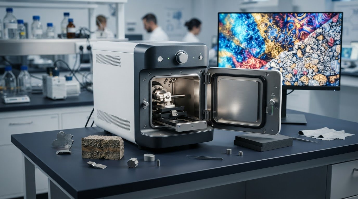
Could a compact SEM deliver the analysis you need, or would its configuration constrain your samples and workflow? The limitations of benchtop scanning electron microscopes aren’t a blanket verdict on compact systems. They depend on what you need to image, the sample’s dimensions and condition, and the capabilities required for routine analysis.
It’s reasonable to ask whether a smaller instrument can accommodate your work. Some constraints apply to SEM analysis broadly; others depend on the particular benchtop model, including its sample capacity, imaging performance, and available detectors or accessories. Separating those factors is essential to making a sound comparison.
This article explains how to assess instrument fit against your sample, imaging goals, and workflow, and when a different SEM configuration may be more suitable. It also outlines how configuration and ongoing support factor into a long-term decision. Electron Optics Instruments, LLC’s service team is available 24/7/365, and its factory-trained service engineers have an average of over two decades of experience.
Key Takeaways
- Assess the limitations of benchtop scanning electron microscopes against your specific sample, imaging objectives, and routine workflow rather than relying on instrument size alone.
- Distinguish considerations shared by SEMs from trade-offs that vary by model and configuration, including specimen dimensions and preparation requirements.
- Use your required outputs and expected sample volume to compare microscopy approaches and determine which configuration best fits your analysis.
- Build a practical selection checklist around representative samples, preparation constraints, throughput, and model-specific documentation.
- Factor product configuration and ongoing support into the ownership decision. EOI’s service team is available 24/7/365, with factory-trained engineers averaging over two decades of experience.
What limitations of benchtop scanning electron microscopes actually mean
A limitation is meaningful only in relation to a defined analytical requirement. A system may be capable of imaging a representative sample for routine inspection yet fall short when another task calls for a larger specimen, finer detail, additional analytical signals, or a different workflow. The limitations of benchtop scanning electron microscopes should therefore be assessed against the work, not inferred from the instrument’s compact footprint.
A benchtop SEM limitation is a boundary between an instrument’s capabilities and the requirements of a specific sample or analysis, not a universal performance threshold. This distinction helps separate general SEM considerations from trade-offs that depend on an individual model and its configuration.
Which limitations are common to SEM, and which depend on the instrument?
Some considerations arise from electron microscopy itself. Conventional SEM workflows generally require samples that can withstand the instrument’s operating conditions. Wet, oily, or outgassing specimens may need preparation or a suitable environmental approach. Nonconductive materials can charge under the beam, although preparation methods and operating modes vary. SEM imaging also describes the surface; viewing internal structure generally requires exposing a cross-section. These principles are part of Scanning Electron Microscope Overview, which introduces SEM components, operation, applications, and limitations.
Other boundaries are model-specific. Chamber dimensions and permissible specimen size, available detectors, operating modes, and achievable imaging performance aren’t identical across benchtop systems. A compact form factor alone doesn’t establish whether a model can accommodate a particular sample or deliver a required analytical output. Those questions depend on the instrument’s documented configuration.
Why the intended application matters more than a general disadvantage list
A general list of disadvantages can obscure the practical decision. A lab conducting routine inspection may need clear surface images and a repeatable process for representative samples. Another workflow may require finer imaging detail, elemental analysis, or examination of specimens with specific preparation challenges. The same benchtop SEM could be appropriate for the first application and unsuitable for the second, depending on its capabilities and the lab’s requirements.
Start with the information the analysis must produce. Is the objective to examine surface features, compare defects, or gather compositional data? Will samples fit the chamber and tolerate the operating conditions? Are preparation steps practical for the team and sample volume? Answering these questions turns broad concerns into requirements that can be evaluated against model documentation. For broader context on beam interaction, image formation, and what SEM can reveal, review the scanning electron microscopy principles that underpin the technique.
- General SEM considerations: specimen condition, preparation, operating environment, and whether the question concerns surface or internal structure.
- Configuration-specific considerations: sample accommodation, available imaging and analytical capabilities, and workflow fit.
This requirements-based view avoids treating “benchtop” as either a guarantee of suitability or an automatic disqualification. Compare the actual specimen, desired outputs, and routine workload with the capabilities of the specific instrument under consideration.
Sample, imaging, and workflow constraints to assess in a benchtop SEM
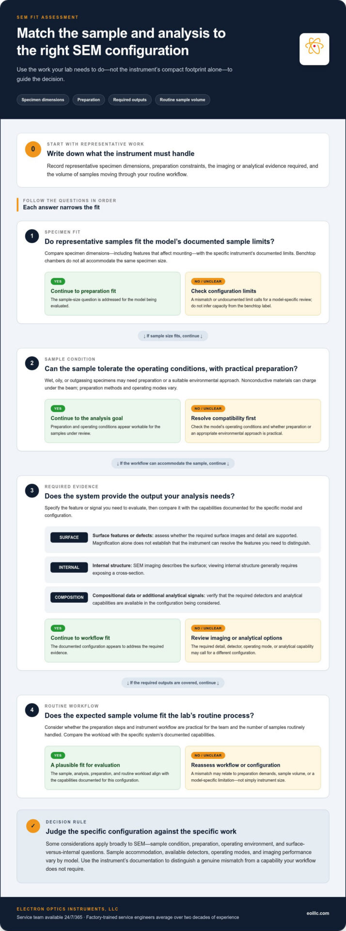
A useful comparison starts with the work the instrument must perform, not a general list of possible drawbacks. Record the specimens you routinely examine, how they must be prepared, what evidence the analysis must provide, and how many samples move through the lab. Before comparing systems, document four variables: specimen dimensions, preparation requirements, required imaging or analytical outputs, and routine sample volume.
These details make the limitations of benchtop scanning electron microscopes more concrete. They also help distinguish a genuine mismatch from a capability your workflow doesn’t need.
Sample dimensions, preparation, and material behavior
Measure representative specimens, including features that affect mounting, and compare those dimensions with the specific instrument’s documented sample limits. Don’t assume all benchtop chambers accommodate the same specimen size. Preparation also depends on the material and operating configuration: conductive and nonconductive samples may call for different preparation steps or imaging approaches. Wet, oily, or outgassing materials require particular attention to compatibility with the instrument’s vacuum conditions. For procedural detail, use a dedicated SEM sample-preparation guide alongside the documentation for the model being considered.
Resolution, analytical needs, and throughput
Magnification and resolution describe different aspects of imaging. A high magnification setting alone doesn’t establish that an instrument can resolve the features your analysis must distinguish. Define the feature of interest and the evidence needed to evaluate it, then compare that requirement with documented performance under relevant operating conditions.
Clarify whether the lab needs surface morphology, topographic contrast, compositional information, or a combination. Imaging and elemental analysis are different requirements, and the latter depends on the system’s configuration. The benchtop SEM options available through Electron Optics Instruments can be evaluated against those requirements, rather than by magnification claims alone.
Throughput matters, too. Estimate the routine number of specimens and include preparation, loading, imaging, analysis, and changeover in the workflow. Compare those steps with the instrument’s documented operating process and your lab’s turnaround expectations. A system that produces appropriate results for occasional investigations may not align with a high-volume inspection workflow if sample handling or analysis becomes a bottleneck.
- Dimensions: Compare actual sample size and mounting needs with the model’s documented limits.
- Preparation: Record material behavior, required preparation, and vacuum compatibility.
- Outputs: Specify whether the task requires morphology, topography, composition, or combined analysis.
- Volume: Account for sample load and the full sequence of preparation and instrument workflow.
Once these requirements are recorded, compare them against model-specific specifications rather than assuming a capability described for one benchtop SEM applies to another. This provides a practical basis for evaluating instrument fit and identifying which constraints matter to your lab.
Benchtop SEM vs. other microscopy options: compare fit, not labels
Choose a microscopy approach by matching the information it provides to the decision your lab needs to make. Optical microscopy may be sufficient when the required observation can be answered with optical images; SEM may be appropriate when electron imaging or additional analytical information is needed. A larger or differently configured SEM is worth evaluating only when documented capabilities of the benchtop options don’t align with the sample or analysis requirements. The limitations of benchtop scanning electron microscopes are relative to the task, not a simple ranking of instrument categories.
Use the comparison below to frame the evaluation. Specimen compatibility and workflow depend on the specific instrument and configuration, so these categories are starting points rather than universal specifications.
| Option | Imaging objective | Specimen requirements | Analysis needs | Sample volume |
|---|---|---|---|---|
| Optical microscopy | Observe features that can be assessed with optical imaging. | Assess whether the specimen and required view are suitable for the optical method. | Use when the decision doesn’t require SEM imaging or SEM-based analytical outputs. | Compare preparation and handling steps with the lab’s routine workload. |
| Benchtop SEM | Examine surface features using electron imaging. | Compare specimen dimensions, condition, and preparation needs with that model’s documentation. | Match imaging and any required compositional analysis to the system’s documented configuration. | Evaluate the documented workflow against expected sample flow and turnaround. |
| Larger or differently configured SEM | Consider when the required imaging task isn’t met by the evaluated benchtop configurations. | Assess when specimen dimensions or conditions exceed a benchtop model’s documented capabilities. | Evaluate when the analysis calls for capabilities or accessories not available in the relevant benchtop configuration. | Compare the complete workflow with the volume and turnaround the lab requires. |
When optical microscopy may meet the imaging requirement
If the question is whether a feature is present in an optical image, or whether samples differ in a way visible through that method, optical microscopy may provide sufficient evidence. If the analysis instead depends on electron imaging or compositional information, assess SEM capabilities against that need. JEOL’s pharmaceutical application example illustrates how benchtop SEM imaging and compositional contrast can address questions beyond a basic visual comparison. It is an application example, not proof that every benchtop SEM suits every pharmaceutical workflow.
When a different SEM configuration may be worth evaluating
Consider another configuration when sample dimensions, required imaging performance, analytical outputs, or routine volume fall outside the documented fit of the benchtop systems under review. Don’t assume a benchtop system cannot perform a task based on its category alone. Compare model-specific specifications and the available analytical setup. For work involving specialized methods, consult an advanced SEM techniques guide to identify which capabilities the analysis actually requires.
The strongest comparison is not “optical versus SEM” or “benchtop versus full-size” in isolation. It is a documented match between the sample, the evidence required, and the workflow the lab must sustain.
A practical checklist for deciding whether a benchtop SEM fits your lab
A structured evaluation makes the choice more defensible than relying on broad claims about benchtop or floor-standing instruments. Start with the analytical decision your lab needs to make, then trace each requirement through sample handling, instrument capability, and ongoing operation. This process turns the limitations of benchtop scanning electron microscopes into specific questions that can be answered with evidence.
- Define the analytical question. State what the analysis must establish, such as whether a surface feature is present, how samples differ, or whether compositional information is required. Separate essential outputs from useful additions so optional capabilities don’t obscure the core requirement.
- Choose representative samples. Record the materials and condition of typical specimens, along with examples that represent the most challenging samples in the routine workload. Include unusual shapes or mounting needs that could affect instrument use.
- Document dimensions and preparation constraints. Note sample measurements, preparation steps, and any material behaviors that may affect imaging or vacuum compatibility. Compare these requirements with the limits and procedures published for each specific model.
- Estimate actual workflow demands. Use your lab’s own sample frequency and turnaround expectations. Account for preparation, loading, imaging, analysis, and changeover rather than considering image acquisition in isolation.
- Identify ownership requirements. Consider operator training, maintenance, replacement parts, and service access as part of the operating plan. Electron Optics Instruments, LLC provides on-site service and training. Its service team is available 24/7/365, and its factory-trained service engineers have an average of over two decades of experience. Plan for maintenance through established SEM maintenance planning as well as day-to-day operation.
- Review model-specific documentation. Match every essential requirement to the exact model and configuration under consideration. Review published specifications for specimen accommodation, imaging performance, analytical capabilities, and workflow, rather than assuming that features or limits transfer between instruments.
The sequence matters. Treat a requirement as a decision criterion only after tying it to a sample or analysis the lab expects to perform. If a capability is essential, identify the corresponding specification or configuration detail in the model documentation. If it’s merely desirable, weigh it accordingly instead of allowing it to drive the selection.
Electron Optics Instruments, LLC’s SEM portfolio includes benchtop and tabletop systems as well as Veritas SEM configurations, providing options to evaluate against different laboratory requirements. Explore EOI’s SEM portfolio as part of a documented comparison of sample fit, analytical needs, and ownership support.
How EOI’s SEM portfolio and on-site support address different lab requirements
Once a lab has defined its samples, required outputs, and workflow, those criteria can guide a focused comparison of instrument models. Electron Optics Instruments, LLC’s portfolio includes benchtop and tabletop SEMs, Genesis systems, and Veritas configurations. Their distinctions provide a starting point for evaluating fit, but model specifications should be considered individually rather than assumed to carry across a product family.
Matching Cube II, Genesis, and Veritas to stated use cases
EOI describes the Cube II SEM as the smallest conventional SEM on the market today. Its stated specifications are 5 nm resolution and magnification up to 300,000×. These figures apply to the Cube II only; they shouldn’t be taken as specifications for other models. Compare them with the lab’s actual imaging requirements and the Cube II’s model documentation.
The Genesis SEM family includes models with different stage configurations. The Genesis-1000 and Genesis-1100 have straightforward, intuitive manual stages. The Genesis-2020 and Genesis-2120 have a more versatile 5-axis motorized stage. The Genesis-1100 also supports observation of non-conductive samples. These distinctions may matter when considering specimen handling and analysis requirements, but they don’t replace a review of model-specific documentation for the complete workflow.
The Veritas SEM is optimized for high-throughput quality-control sample analysis and requires no complex utilities for operation. That stated focus can inform the shortlist for a lab assessing routine inspection; the decision should still reflect the lab’s analytical outputs, sample demands, and operating context. Across the portfolio, the practical question isn’t which family sounds most capable in general, but which documented configuration aligns with the required work.
On-site service as part of the instrument decision
Instrument fit extends beyond image acquisition. Training, maintenance, parts, and service access all affect how a system is incorporated into lab operations and supported over its equipment lifetime. EOI engineers provide service and training on site, helping labs plan for continued operation as part of a lifetime support relationship.
EOI’s factory-trained service engineers have an average of over two decades of experience, and the service team is available 24/7/365. EOI provides preventative maintenance, repairs, on-site training, and SEM relocation and site evaluation. Its engineers perform service and training on site, so there is never a need to send your SEM back to the manufacturer’s facility. Include support considerations in the same evaluation as sample fit and analytical capability, particularly when uptime and staff readiness matter to the lab’s routine.
The limitations of benchtop scanning electron microscopes become clearer when each model is assessed against specific requirements rather than category assumptions. To discuss which EOI SEM configuration and on-site support approach align with your lab’s needs, contact Electron Optics Instruments, LLC.
Turn your lab requirements into a confident SEM decision
The most useful next step is to turn your evaluation into a shared specification: identify the samples and outputs that matter most, document the non-negotiable requirements, and use those criteria to guide instrument selection. That gives your team a clear basis for discussing where the limitations of benchtop scanning electron microscopes matter in practice and where a compact configuration may fit the work.
Instrument choice also shapes the workflow that follows. Consider how your selected system will support the people operating it, the analyses your lab expects to perform, and the way equipment support fits into long-term planning. A requirements-led discussion can connect those priorities to an appropriate SEM configuration and support approach.
Discuss your SEM requirements with EOI to explore instrument fit and support for your lab. With a clear set of priorities, you can move forward with greater confidence and build a microscopy workflow aligned with your analytical goals.
Frequently Asked Questions
What is the main limitation of a benchtop scanning electron microscope?
There isn’t one limitation shared by every benchtop model; the key question is whether a specific configuration can accommodate your specimen and provide the evidence your analysis requires. A sample might fit physically but still need preparation or analytical capabilities that the selected setup doesn’t support. Consider the limitations of benchtop scanning electron microscopes in relation to your actual samples and decisions, including whether a mismatch could affect result quality or disrupt routine work.
Can a benchtop SEM analyze non-conductive samples?
Yes, some models support observation of non-conductive samples, but that capability shouldn’t be assumed across all benchtop SEMs. For example, the Genesis-1100 is specified for observing non-conductive samples. Before planning a workflow, consider the material, the image or analysis required, and the preparation or operating approach described for the selected configuration. This helps determine whether a particular instrument is suitable for your specimens without generalizing from one model to another.
How do I know if my sample is too large for a benchtop SEM?
Compare the specimen’s complete mounted dimensions with the selected SEM’s documented sample limits, not just the size of the unmounted material. Include any holder or fixture that will remain attached during imaging, and account for the orientation needed to examine the target area. If a specimen can be prepared as a smaller representative section, consider whether that changes the analytical question. The instrument documentation should guide the final fit assessment.
Is a benchtop SEM suitable for quality-control work?
It can be, if the system’s documented capabilities and workflow align with the inspection task. EOI describes its Veritas SEM as optimized for high-throughput quality-control sample analysis, with no complex utilities required for operation. For a practical assessment, define the feature inspectors must identify, the records or outputs required, and how samples move through the process. Then compare those needs with the specific configuration. A quality-control label alone doesn’t establish suitability for every inspection protocol.
When should a lab consider a larger or differently configured SEM?
Evaluate another configuration if a required specimen, analytical output, or operating workflow falls outside the published capabilities of the benchtop models under consideration. For instance, a project involving a specialized analytical method may require a configuration with particular instrumentation. First identify the method and its requirements, then compare documentation for suitable systems. A larger instrument isn’t automatically the better choice; the deciding factor is whether its capabilities address a defined need.
Does a benchtop SEM replace optical microscopy?
No, not as a universal replacement. The methods provide different information, so the appropriate choice depends on the observation required. If an optical image can answer a visual inspection question, SEM may add complexity without serving the immediate purpose. If the task calls for electron imaging or SEM-based compositional information, optical imaging alone may not answer it. Some workflows use both methods at different stages, with each contributing evidence suited to its purpose.
Can EOI service an SEM that was purchased from another supplier?
Yes. Electron Optics Instruments provides service for SEMs from other major manufacturers as well as equipment sold through EOI. Support is arranged through a service contract, with an initial consultation to establish the instrument and support requirements. EOI’s service team is available 24/7/365, and its factory-trained service engineers have an average of over two decades of experience. This gives labs a way to plan service for equipment already in operation, taking its manufacturer and condition into account as part of the service arrangement.