
More magnification can make an SEM image look larger without revealing another specimen feature. If fine details remain soft as you zoom in, the cause may lie in the instrument, the sample, or the imaging conditions, not in the magnification setting alone. Maximizing SEM imaging resolution means understanding how these factors work together and improving useful detail without sacrificing interpretability.
Magnification enlarges the image, while resolution determines whether closely spaced features appear as distinct details. For example, the Cube II SEM has a specified resolution of 5 nm and magnification of up to 300,000×. These figures describe different capabilities, and one does not guarantee the other.
This article explains what affects SEM resolution and gives you a repeatable workflow for assessing image detail. You’ll learn how beam conditions, working distance, sample characteristics, preparation, and instrument performance can influence results, and how to distinguish operating issues from a possible instrument-performance concern.
Key Takeaways
- For maximizing sem imaging resolution, define the feature you need to resolve and save a baseline image before changing settings.
- Adjust one imaging condition at a time so you can identify which changes improve useful detail and which introduce artifacts.
- Assess image quality by the specimen detail you can distinguish, not by image size alone.
- Consider instrument performance, specimen response, and imaging conditions together when diagnosing soft or unclear images.
- EOI supports SEM operation through service and training. Its factory-trained service engineers average over two decades of experience, and the service team is available 24/7/365.
What SEM Imaging Resolution Actually Measures, and Why More Magnification Is Not Enough
You increase the image size, expecting to see another level of specimen detail, but the result is simply a larger blur. This is a common frustration in SEM imaging: magnification changes how large the image appears, but it cannot reveal information the system has not resolved.
SEM imaging resolution is the ability to distinguish closely spaced specimen features as separate details in an image. It describes how clearly information can be separated, not the size at which that information is displayed. The Scanning Electron Microscope overview explains the instrument’s operating principles and components, which help show why image detail depends on more than magnification alone.
How to tell resolution apart from magnification
Magnification enlarges the displayed image; resolution determines whether nearby features remain distinguishable as separate structures. Imagine two adjacent ridges on a specimen. If the image shows two distinct ridges with a visible boundary between them, their detail is resolved. If they merge into one indistinct area, increasing magnification enlarges that merged appearance without separating the ridges.
An enlarged image can look more prominent without containing more specimen information. A feature that appears blurred at one display size remains unresolved if the image does not distinguish its boundaries. Enlarging it may make the blur easier to inspect, but it will not create a clear separation that was not captured.
Judge an image by whether it answers the question you set out to investigate: can you identify the feature, distinguish it from neighboring structures, and interpret its boundaries? The displayed scale is useful, but it does not prove by itself that finer detail has been captured.
Why useful resolution depends on the imaging task
The required level of detail depends on the analytical question. If you need to assess broad morphology, such as a specimen’s overall shape, distribution, or surface condition, a clear view of larger structures may be enough. If you need to distinguish fine surface features from their immediate surroundings, the image must preserve more localized detail.
There is no single resolution target that suits every specimen or application. A setting that provides useful information for a broad survey may not answer a question about small surface structures. Pursuing finer detail than the task requires can also add effort without improving interpretation. Define the feature of interest first, then assess whether the image separates it from nearby features.
This task-led approach is central to maximizing sem imaging resolution: aim for interpretable detail that supports the analysis, not the largest possible display. Distinguishing enlargement from resolved information also makes it easier to compare images and choose adjustments that could meaningfully improve them.
Which SEM Imaging Conditions Influence the Detail You Can Resolve?
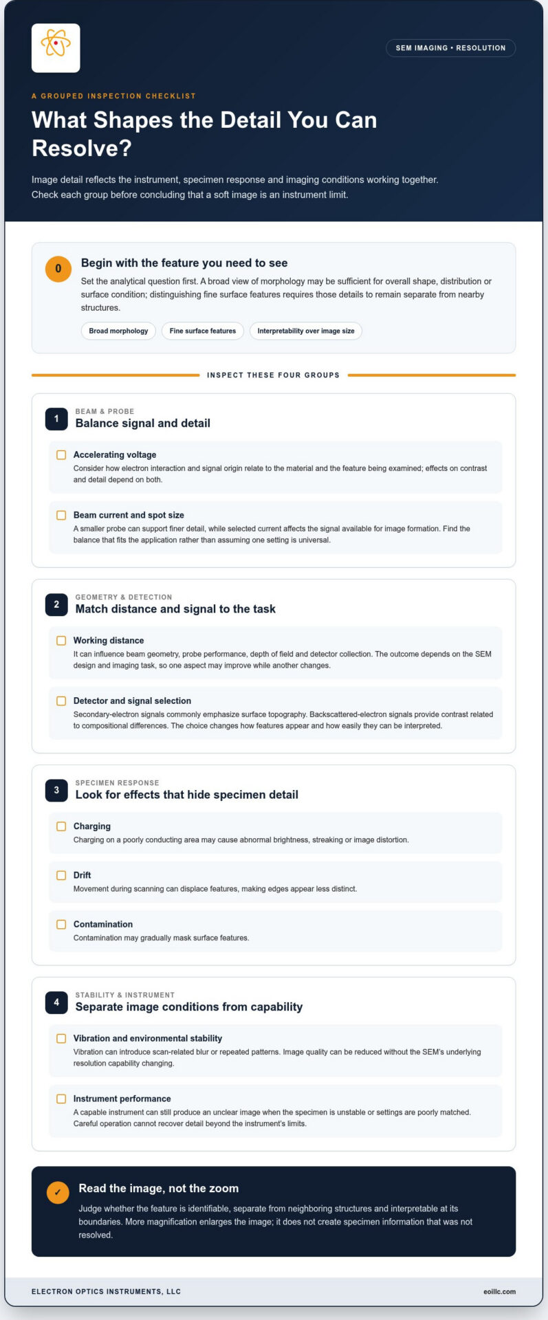
A soft image does not automatically mean the SEM has reached its resolution limit. The detail captured in a scan reflects the instrument’s capability, the specimen’s response to the electron beam, and the imaging conditions. A capable instrument can produce an unclear image if the specimen is unstable or the settings are poorly matched to the task. Careful operation, in turn, cannot recover detail beyond the instrument’s limits.
No single SEM setting guarantees the best-resolved image. Beam energy, probe conditions, working distance, signal selection, specimen behavior, and environmental stability interact with the instrument and the imaging task. Assessing these factors together helps distinguish a correctable imaging issue from a potential instrument-performance concern.
Beam conditions, working distance, and signal selection
Accelerating voltage affects how electrons interact with a specimen and where detected signals originate. Its influence on contrast and detail depends on the material and the feature being examined. Probe conditions, including beam current and spot size, also involve trade-offs. A smaller probe can support finer detail, while the selected current affects the signal available for image formation. The right balance depends on the application, not on a universal setting.
Working distance affects the beam’s geometry at the specimen and can influence probe performance, depth of field, and detector collection. The result depends on the SEM design and the imaging task, so changing working distance may improve one aspect of an image while altering another. Detector choice matters too: secondary-electron signals commonly emphasize surface topography, while backscattered-electron signals provide contrast related to compositional differences. Signal selection affects how features appear and can change interpretability, even when the specimen is unchanged.
Sample behavior and environmental stability
Specimen behavior can obscure detail the instrument could otherwise capture. Charging on a poorly conducting area may cause abnormal brightness, streaking, or image distortion. Drift can move features during scanning, making edges appear displaced or less distinct. Contamination may gradually mask surface features, while vibration can introduce scan-related blur or repeated patterns. These effects reduce image quality, but do not necessarily indicate that the SEM’s underlying resolution capability has changed.
Look for patterns in the image. Distortion that varies across a specimen or changes over time may point to charging, drift, contamination, or instability rather than a fixed resolution limit. Compare areas and scans under consistent conditions before drawing conclusions. Sample properties and preparation affect how a specimen behaves under the beam, so consult an SEM sample preparation guide for preparation-specific guidance.
To maximize SEM imaging resolution, separate what the instrument can resolve from what the specimen and operating environment allow you to see in a particular scan. If the intended feature remains indistinct after systematic adjustments, the question may shift from image optimization to instrument performance. EOI’s SEM imaging systems provide a starting point when considering equipment for your imaging requirements.
Common Resolution Misconceptions: How to Judge SEM Image Quality Correctly
SEM images can appear more impressive after enlargement or contrast adjustment, but appearance alone does not establish how much specimen detail has been resolved. Reliable interpretation depends on separating image scale, visible contrast, edge sharpness, and the ability to distinguish neighboring features. These distinctions help prevent a visually striking image from being mistaken for a more informative one.
| Misconception | Reality |
|---|---|
| Higher magnification always reveals finer detail. | Magnification enlarges the displayed image; it cannot recover detail the imaging system did not resolve. |
| A sharp-looking image necessarily has high resolution. | Sharpness describes how crisp edges appear. Resolution concerns whether separate nearby features can be distinguished. |
| More contrast means better resolution. | Contrast makes differences easier to see, but it does not necessarily separate features that merge together. |
| A stated resolution specification guarantees the same detail on every sample. | It describes instrument capability under defined conditions; specimen behavior and operating conditions affect the image obtained. |
Does higher magnification always produce higher resolution?
No. Magnification alone cannot recover detail the imaging system has not resolved. If two neighboring features are recorded as one blurred region, enlarging that region makes the blur more conspicuous, not the features more distinct. At high enlargement, image pixels or scan-related softness may also become easier to see, creating an impression of added detail without revealing new specimen information.
Choose magnification in relation to the feature you are evaluating and the image’s ability to show its boundaries. For a broad surface pattern, a wider field may provide the context needed for interpretation. For a small feature, increase magnification only while the image continues to show meaningful separation, not just a larger, less clear view.
How to interpret a stated SEM resolution specification
A manufacturer’s resolution specification is best understood as a capability measured under particular test conditions, not a promise that every sample will show the same level of detail. Specimen properties, beam conditions, signal selection, and image stability can all affect practical results. Compare specifications only when their definitions, test samples, and measurement conditions are sufficiently comparable.
SEM resolution measurement methods are not universally standardized across manufacturers, making headline figures difficult to compare in isolation. Treat a specification as one part of equipment assessment, then consider whether the SEM can deliver useful detail for the actual analytical task. For maximizing sem imaging resolution, judge the result by interpretable features, not magnification, contrast, or a specification number alone.
In practice, assess edge definition, separation of neighboring structures, and consistency across comparable scans. A crisp boundary may improve visual clarity, while strong contrast can make a feature easier to locate. Neither necessarily proves that closer features are separately resolved. Keeping these image-quality measures distinct supports disciplined interpretation and more meaningful comparisons between images.
A Repeatable Workflow for Maximizing SEM Imaging Resolution
Improving image detail is more reliable when you treat each adjustment as a controlled test rather than changing several settings at once. Start with the feature you need to examine, establish a stable baseline, and record what changes. Controlled, one-at-a-time adjustments make SEM troubleshooting more reliable because they show which change affects visible detail.
Use this sequence to assess image quality and preserve a useful reference for comparison:
- Define the target feature. Identify the structure or boundary the image must reveal, and select a representative sample area. Decide what would count as a meaningful improvement, such as clearer separation between neighboring features, rather than simply a larger display.
- Confirm stable imaging conditions. Check whether the specimen is drifting or charging, and verify that focus and astigmatism are not making edges appear soft. If the image changes over time or the specimen moves during scanning, address that instability before judging fine detail.
- Record a baseline. Note the sample area, magnification, working distance, signal or detector, and current acquisition conditions. Save the image with these settings so you can compare it with later scans. Follow your SEM operation guide for instrument-specific procedures and controls.
- Optimize focus and astigmatism. Bring the feature of interest into focus, then correct astigmatism according to the instrument’s operating procedure. Recheck the target feature after each adjustment. Poor focus or astigmatism can distort edges, so assessing resolution before correcting them can lead to misleading conclusions.
- Change one relevant condition at a time. Adjust a single acquisition variable, such as beam conditions, working distance, or signal selection, while keeping the target area and other recorded conditions consistent. Save the new image and note whether the feature is more distinctly separated, less distinct, or unchanged.
- Compare like-for-like images. Review the baseline and adjusted scans at the same display scale and evaluate the same specimen area. Look for reproducible improvement in the detail relevant to your task, not just greater contrast or a sharper-looking edge.
- Document persistent problems. If results remain inconsistent after controlled adjustments, preserve representative images and settings, and record symptoms such as drift, charging, or changes in focus. This information gives service personnel a clearer basis for assessing instrument performance.
Maximizing sem imaging resolution is a systematic process, not a search for one universally optimal setting. A record of settings and image outcomes helps separate a condition-specific issue from a repeatable performance concern.
If image quality remains inconsistent, EOI provides service support to its purchasers and third-party SEM owners through service contracts. Its factory-trained service engineers average over two decades of experience, and the service team is available 24/7/365. EOI engineers perform service on site. Explore SEM service support for help with ongoing instrument operation.
How EOI Supports SEM Resolution and Reliable Imaging Performance
Clear SEM results depend on more than a resolution figure. Instrument capability, specimen behavior, operating conditions, and consistent system performance all influence the detail an operator can interpret. EOI supports this work through SEM equipment, service, maintenance, and training, helping operators build reliable practices and maintain their equipment.
Match SEM capability to the imaging requirement
Choosing an SEM begins with the features and analytical questions your imaging process needs to address. The Cube II SEM is a tabletop SEM with a specified 5 nm resolution and magnification of up to 300,000×. These are separate specifications: resolution describes the stated capability to distinguish detail, while magnification describes image enlargement. Neither number alone establishes the detail every specimen will show under every condition.
Consider the required field of view, specimen characteristics, and whether the task involves broad morphology or finer surface structures. EOI offers benchtop and tabletop SEMs for a range of imaging requirements. A broader understanding of desktop SEM configurations can help put equipment choices in context. Matching capability to the task is a more practical starting point than selecting by maximum magnification alone.
Maintain performance through experienced service and training
Consistent operation also depends on appropriate maintenance and informed use. EOI’s factory-trained service engineers average over two decades of experience, and the service team is available 24/7/365. EOI provides service support to its purchasers and third-party SEM owners through service contracts. The company’s engineers perform service and training on site.
On-site training can help operators apply instrument procedures consistently, while Full Preventative Maintenance Service Contracts support ongoing equipment care. These resources support operational reliability and repeatable imaging practices. They do not guarantee a specific resolution or image-quality result.
For maximizing sem imaging resolution, treat the instrument specification as one part of a wider performance picture. If image detail changes unexpectedly, recorded operating conditions and specimen behavior can help distinguish a routine adjustment issue from a concern that warrants service assessment. EOI’s SEM service, maintenance, and training support this broader approach to reliable imaging performance.
Explore EOI’s SEM equipment and service support to consider options for your imaging and operational requirements.
Make Clear Imaging a Repeatable Part of Your Workflow
Translate image-quality goals into practical criteria your team can apply across sessions. Identify the specimen features that must be interpretable for the analysis, then agree on how images will be reviewed and documented. This gives operators a shared reference for deciding whether an image is fit for its purpose, rather than relying on individual impressions of sharpness or display size.
As imaging needs evolve, revisit those criteria alongside your SEM capabilities, operating practices, and maintenance planning. A consistent framework connects day-to-day image evaluation with longer-term equipment decisions, while leaving room to adapt methods to new specimens and analytical questions. That is a practical foundation for maximizing sem imaging resolution: progress is measured by dependable, useful information, not enlargement alone.
For SEM equipment, preventative maintenance, on-site service, or training, explore EOI SEM equipment and service support to discuss your imaging workflow.
Frequently Asked Questions
What does resolution mean in an SEM?
SEM resolution describes whether nearby specimen features appear as distinct details rather than merging together. For example, in a quality-control image of a coated surface, the useful question may be whether adjacent defects can be identified separately, not how large they appear on screen. Set the required detail according to the analysis, then judge whether the image supports that decision. Magnification changes image scale, but it cannot substitute for resolved information.
Can increasing SEM magnification improve resolution?
Not on its own. Increasing magnification may help you inspect a feature that is already distinguishable, but it cannot create a separation absent from the captured image. If a boundary looks soft, first check whether the feature is in focus and whether the specimen is stable. Compare images of the same area at consistent display scales. This helps you avoid treating a larger view as proof that finer detail has emerged.
What factors affect SEM imaging resolution?
Image detail reflects the instrument, its operating conditions, and how the specimen responds during imaging. Beam settings and working distance can affect signal formation, while detector selection influences which specimen characteristics stand out. Focus, astigmatism, charging, drift, contamination, and vibration may also reduce apparent detail. Their impact varies with the specimen and purpose of the scan, so record conditions and investigate changes in a controlled way rather than relying on a universal setting.
Does pixel size determine SEM resolution?
No. Pixel size describes how the image is sampled, not whether the microscope distinguished the specimen features in the first place. If sampling is too coarse, small image details may be represented poorly. Finer sampling can improve how captured information is displayed, but it cannot recover detail that was not recorded. Assess pixel sampling alongside focus, signal quality, scan conditions, and the feature’s appearance in the original acquisition.
How can I tell whether an SEM image is truly sharper?
Compare images of the same specimen area using documented, comparable acquisition conditions. Look for a feature boundary or pair of neighboring structures that becomes consistently easier to distinguish, rather than relying on a brighter image or stronger contrast. Keep display scale and image processing consistent during comparison. If the apparent improvement disappears when these factors are matched, it may reflect presentation changes rather than a meaningful increase in visible specimen detail.
Why does my SEM image look blurry at high magnification?
At high magnification, small focus errors, astigmatism, specimen movement, charging, vibration, or weak signal can become more apparent. Start by checking whether the image changes over time or whether edges look soft in every direction. Recheck focus and astigmatism, then compare a stable scan of the same area. If blurring persists across controlled checks, preserve the images and settings so service personnel can assess the pattern.
How often should SEM resolution be checked?
Set a verification schedule that fits the instrument documentation, laboratory quality procedures, application, and frequency of use. Keep reference images of a suitable specimen or feature, captured under repeatable conditions, so changes can be recognized over time. Reassess after maintenance, a change in imaging setup, or an unexpected shift in image quality. EOI offers Full Preventative Maintenance Service Contracts as one option for supporting planned SEM upkeep.