
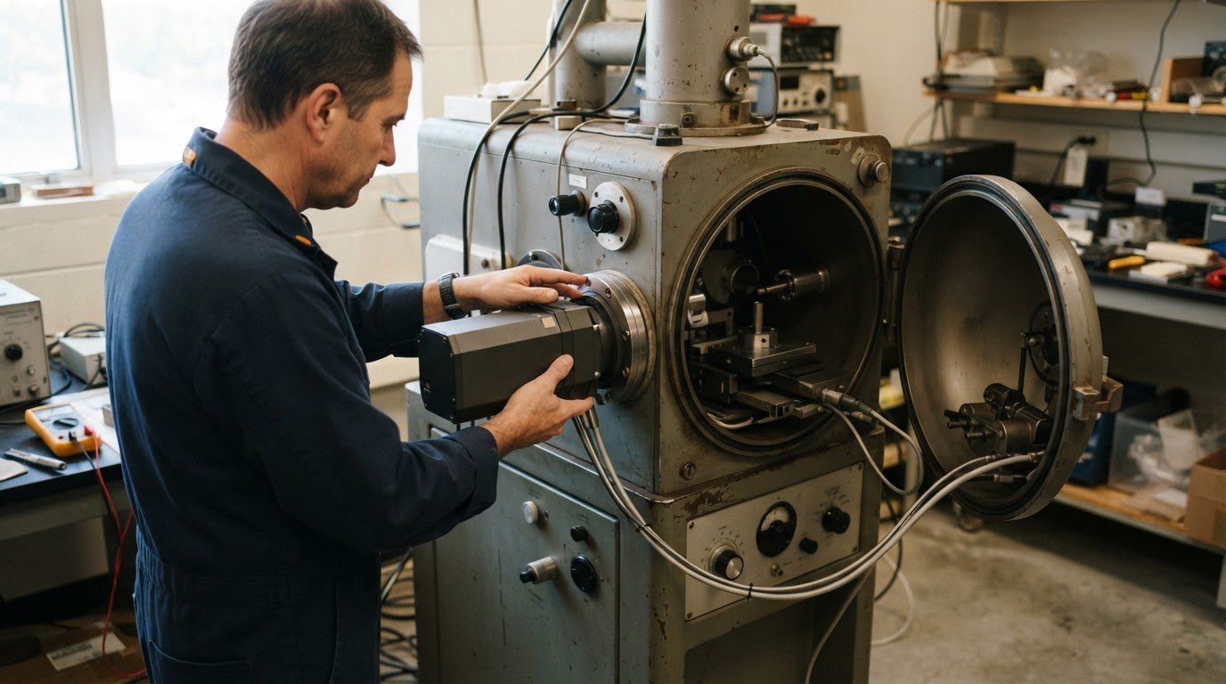
An older SEM doesn’t automatically rule out elemental analysis. In many cases, you can add EDS to an old SEM if its configuration supports the detector and software integration. EDS measures X-rays emitted from a specimen under an electron beam, helping identify elements and show where they are distributed.
It’s reasonable to ask whether an upgrade will work with your existing instrument and how installation will affect established workflows. Feasibility depends on practical compatibility, including the microscope’s design, available detector space, and integration of the system’s hardware and software.
This guide explains how EDS extends SEM imaging with qualitative and quantitative materials analysis, what affects upgrade feasibility, and what installation and operator training involve. It also covers detector integration, system configuration, and analysis workflows, so you can assess the technical considerations before planning an upgrade.
Key Takeaways
- An EDS upgrade can extend an older SEM’s capabilities, but compatibility depends on the instrument’s configuration and how detector and software integration can be achieved.
- Learn how EDS supports elemental identification, composition estimates, and chemical mapping, and what these results can contribute to materials analysis.
- Understand the main stages of installation and training, from integrating the detector and software to preparing operators to interpret EDS data.
- Explore the operating and maintenance considerations that help determine whether retrofitting is a practical path for your SEM.
Upgrade installation and training
An EDS retrofit adds an X-ray detector and analysis capability to an SEM workflow. Installation connects the detector and software to the microscope, while training helps operators interpret elemental data alongside images.
Compatible SEM Models
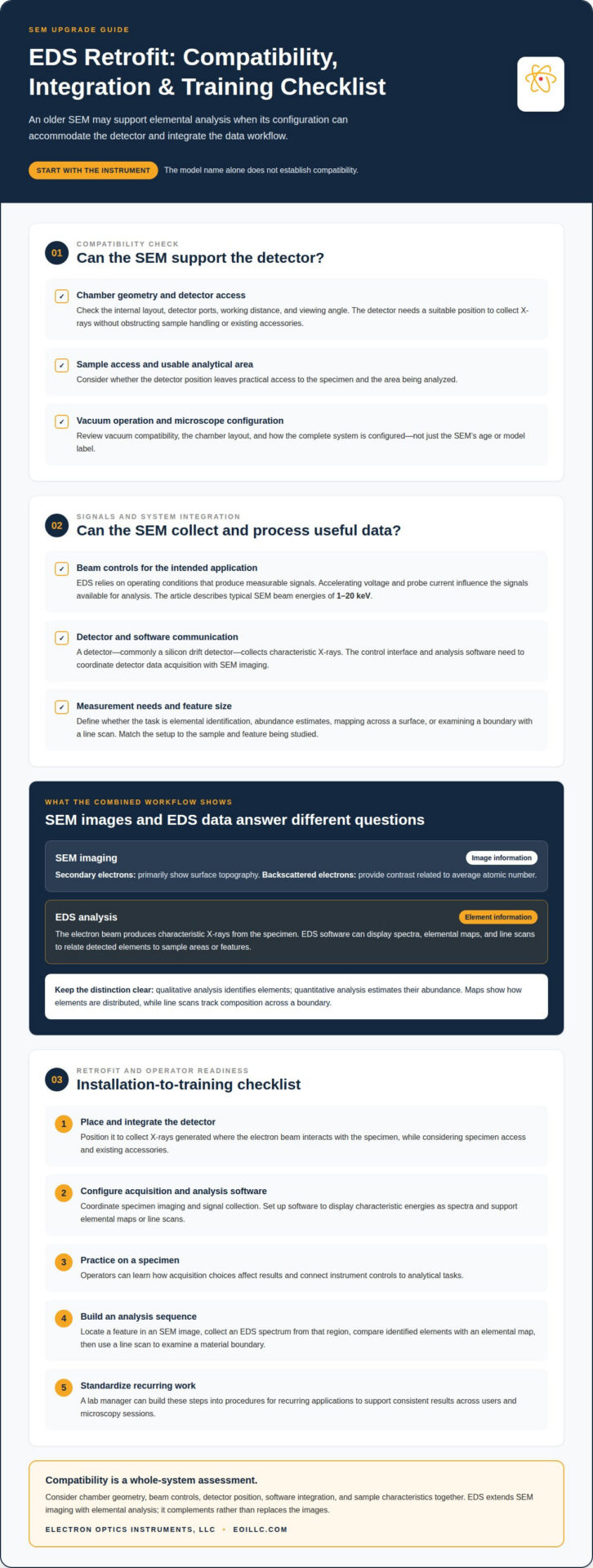
EDS compatibility depends on whether an SEM can accommodate a detector and integrate its signals with suitable analysis software. The model name alone does not establish compatibility. Chamber geometry, system configuration, and software integration all affect whether the detector can collect useful X-rays while the electron beam scans a specimen.
As the electron beam interacts with a sample, it generates characteristic X-rays. An EDS detector, commonly a silicon drift detector, collects these X-rays for software analysis. The resulting elemental information adds chemical analysis to SEM imaging: secondary electrons primarily show surface topography, while backscattered electrons provide contrast related to average atomic number.
Compatibility also shapes which applications an upgraded instrument can support. EDS software can display spectra, elemental maps, and line scans, helping operators relate detected elements to specific areas or features. A map can show how elements are distributed across a surface, while a line scan can track composition across a boundary. These outputs complement SEM images rather than replace them.
Retrofit Installation and Training
EDS installation integrates the detector with the SEM and configures the software used to acquire and interpret X-ray data. Reliable analysis depends on coordinating specimen imaging, signal collection, and the selected measurement workflow, not simply adding hardware to the microscope.
Installation centers on detector placement, system integration, and software setup. The detector must collect X-rays generated where the electron beam interacts with the specimen. The software displays characteristic energies as a spectrum and supports mapping or line scans. After integration, operators can use a specimen to learn how acquisition choices affect the results.
Training should connect instrument controls to analytical tasks. Operators learn to recognize elements from characteristic X-ray signals, review spectra, and create maps or line scans for relevant sample areas. They also need to distinguish qualitative identification from quantitative estimates of elemental abundance, since these results answer different questions.
A practical training exercise might start by locating a feature in an SEM image and collecting an EDS spectrum from that region. The operator can compare the identified elements with an elemental map, then use a line scan to examine a material boundary. A lab manager can build these steps into procedures for recurring applications, supporting consistent results across users and microscopy sessions.
What SEM models are compatible with EDS detectors?
EDS detectors can be integrated with SEMs when the instrument’s configuration supports detector placement, signal collection, and software communication. The microscope’s age alone doesn’t determine compatibility. Whether you can add EDS to an old SEM depends on the chamber layout, detector access, vacuum operation, and how the control system handles analytical data.
Start with the chamber and specimen stage. An EDS detector needs a suitable position to collect X-rays without obstructing sample handling or existing accessories. The chamber’s internal layout, working distance, and detector ports affect its viewing angle and usable analytical area. A compact tabletop SEM and a larger floor-standing SEM may therefore require different integration approaches.
Beam control is another consideration. A typical SEM raster-scans a focused electron beam across a sample at energies of 1 to 20 keV. EDS analysis depends on the operating conditions that produce measurable signals, so the microscope’s beam controls must support the laboratory’s intended applications. Accelerating voltage and probe current influence the signals available for analysis.
Detector technology and software also matter. A silicon drift detector collects X-rays, while analysis software processes the signal into spectra and elemental maps. The SEM’s control interface needs to coordinate detector data acquisition with imaging. To decide whether you can add EDS to an old SEM, consider whether its architecture can connect these functions into a workable analytical process.
Application requirements help define compatibility in practical terms. Qualitative analysis identifies elements, while quantitative analysis estimates their abundance. Mapping can reveal how elements vary across a surface, and a line scan can examine a material boundary. Feature size matters when matching an instrument and detector to a measurement task.
Assess the complete instrument rather than relying on its model label alone. Chamber geometry, beam controls, detector position, software interface, and sample characteristics all shape whether an EDS setup can provide useful results. A system intended to locate a broad contaminant patch, for example, has different analytical needs from one examining a narrow material interface.
For an instrument review, SEM and EDS system options provide a relevant starting point for considering detector integration alongside microscope configuration.
What safety, maintenance, and operating risks come with retrofitting EDS?
Retrofitting EDS changes the SEM chamber configuration, software workflow, and routine operating requirements. Before installation, the integration plan should account for detector placement, specimen access, vacuum compatibility, and existing microscope controls. For example, a detector positioned too close to a holder or accessory can restrict access to the sample area.
Installation involves working with an instrument designed to operate under vacuum. The chamber must be opened and returned to operating conditions, so trained personnel should follow the SEM’s service procedures. After installation, check vacuum operation and detector communication before analytical work resumes. This helps identify integration problems before they affect routine imaging or data collection.
Sample conditions can create analytical challenges even when the hardware works correctly. Insulating specimens may need coating for SEM imaging, while carbon on a specimen surface can complicate EDS interpretation because it may be contamination rather than part of the material. Coatings and surface deposits can influence the spectrum, so operators should account for sample preparation and surface condition when interpreting elemental results.
EDS data require careful interpretation. A spectrum can identify elements, while quantitative analysis estimates their abundance; these results are not interchangeable. Elemental maps and line scans show spatial patterns, but a map may reflect the chosen acquisition conditions as well as the specimen. A signal near a material boundary, for instance, should be interpreted in context rather than treated as a definitive description of the entire sample.
Routine care supports detector performance and SEM operation. Keep the detector’s operating environment clean, follow the instrument’s maintenance procedures, and address changes in signal or software communication before they disrupt analysis. Preventative Maintenance Visits can support planned SEM upkeep, particularly when an EDS upgrade adds another component to the operating workflow.
Operating procedures should define who can change acquisition settings, how data are named and stored, and how results are reviewed. A lab manager can establish a consistent workflow for recording sample identity, imaging conditions, and analytical outputs. This supports traceability when different operators examine similar specimens and makes it easier to compare spectra or maps across sessions.
For help maintaining SEM performance after an upgrade, schedule a Preventative Maintenance Visit as part of your instrument-care plan.
Electron Optics Instruments
Electron Optics Instruments helps laboratories evaluate SEM options when elemental analysis is part of a broader microscopy plan. If an existing instrument’s configuration limits the required workflow, considering another SEM may be a practical next step alongside assessing retrofit feasibility.
Its SEM offerings include the Cube II Benchtop SEM, Genesis Tabletop SEM, Veritas Series SEM, and Veritas Plus SEM. The right system depends on the samples, imaging requirements, and analytical tasks the laboratory needs to support. Clarifying those priorities helps frame a useful discussion about instrument direction without assuming every SEM suits every application.
If you’re assessing an older SEM or considering another system for EDS-related work, contact Electron Optics Instruments to discuss your requirements.
Plan the next stage of your SEM workflow
Whether you can add EDS to an old SEM depends on how its configuration supports detector integration and meets your laboratory’s analytical needs. That assessment can also help determine whether extending the current instrument or considering another SEM is the better fit for future work.
Electron Optics Instruments offers the Cube II Benchtop SEM, Genesis Tabletop SEM, and Veritas Series SEM. These systems provide options to consider when an existing instrument no longer meets the lab’s microscopy requirements. A discussion of your samples, imaging priorities, and elemental-analysis goals can help clarify the direction that best fits your work.
Share your SEM and application requirements with Electron Optics Instruments to discuss the next step. Contact Electron Optics Instruments to begin planning with a clear view of your instrument needs and analytical objectives.
Frequently Asked Questions
Can EDS be added to an older scanning electron microscope?
Yes, an older SEM can accept EDS when its chamber, controls, and software support detector integration. Age alone doesn’t determine whether an upgrade is practical. A microscope with suitable detector access and a compatible data interface, for example, may support EDS without replacing the entire system. The key factors are the SEM’s configuration and condition, not simply its manufacturing date.
What components are needed to retrofit EDS to an SEM?
A typical EDS retrofit includes an X-ray detector, a mounting arrangement, analysis software, and connections for exchanging data with the SEM. The detector collects X-rays generated from the specimen, while software processes them into spectra and other analytical outputs. Installation also needs to account for the chamber layout and how operators select image areas for analysis.
Can a used or refurbished EDS system be installed on an older SEM?
A used or refurbished EDS system can be installed if its detector and software integrate with the SEM’s configuration. Compatibility involves more than physical fit: the system also needs functioning electronics, suitable data connections, and software that communicates with the microscope. An older detector with incompatible control software, for example, may not support the intended analytical workflow without further integration.
Does an older SEM need upgrades to its vacuum, electronics, or software for EDS?
Not every older SEM needs changes to all three systems. Vacuum requirements depend on the microscope’s operating condition and sample workflow, while electronics and software determine whether detector signals can be acquired and interpreted. An SEM may image normally but need a software or interface upgrade to coordinate EDS data collection with image acquisition.
What are the installation and calibration steps for adding EDS to an SEM?
The process generally involves fitting the detector, connecting it to the SEM, configuring the analysis software, and testing data acquisition. Calibration checks whether the detector’s energy scale and spectral response support reliable interpretation. A practical validation uses a known reference material, followed by a test acquisition to confirm that the system produces expected spectral features before routine sample analysis begins.
Who provides EDS retrofit services and support for older SEMs?
Electron Optics Instruments provides EDS systems and upgrades older EDS X-ray systems to Windows-based software. It also services and repairs electron microscopes from major manufacturers. If your laboratory is considering EDS for an older instrument, contact the company to discuss your equipment and analysis needs. Its SEM options include the Cube II Benchtop SEM, Genesis Tabletop SEM, and Veritas Series SEM.