
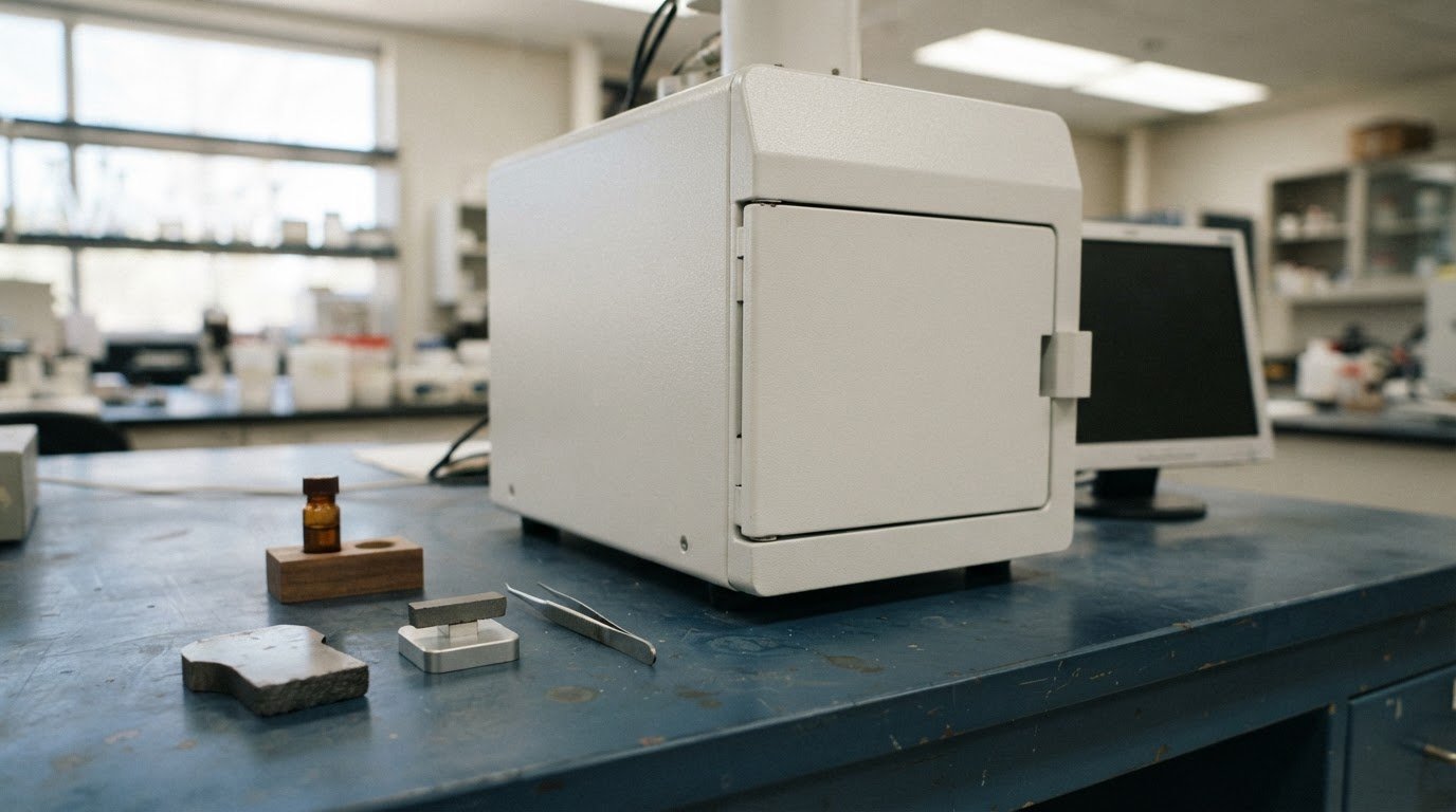
A benchtop SEM can do more than produce surface images. Depending on its configuration, it can support EDS for elemental analysis or EBSD for crystallographic characterization. Advances in benchtop SEM technology have also expanded imaging options, including low-vacuum operation for some nonconductive or beam-sensitive samples. The right setup depends on the material and the information you need.
Adding an analytical technique involves more than attaching a detector: compatibility, sample characteristics, and the data you need all shape the system. This overview explains how low- and high-vacuum imaging differ, what benchtop SEMs can contribute to materials characterization, and how EDS or EBSD can extend analysis. It also looks at how a more accessible SEM platform can bring imaging and characterization into routine lab workflows, and when a benchtop system suits the task.
Key Takeaways
- Assess sample dimensions, conductivity, and beam sensitivity to determine which benchtop SEM setup suits your materials analysis.
- Understand how low-vacuum and high-vacuum imaging affect the way different samples can be examined.
- Identify when EDS or EBSD may add useful elemental or crystallographic information to SEM imaging.
- Explore how a benchtop system, including the Cube II Benchtop SEM, could support analysis within your laboratory workflow.
Can you give us an example of a case where the desktop SEM was particularly useful?
A desktop SEM is particularly useful when a materials team needs surface images from a component without sending it elsewhere. For example, an engineer examining a fractured coating can locate cracks and compare damaged edges with intact regions. Newer benchtop SEMs can accommodate samples up to 70 mm in diameter and 50 mm thick, allowing examination of larger specimens than earlier units, which accommodated samples up to 25 millimeters (mm) in diameter.
Start sample preparation by considering the material and the question you want to answer. A conductive specimen may need minimal preparation, while a nonconductive sample can charge under the electron beam and affect the resulting images. Ion beam milling can expose a clean cross-section or refine a surface before imaging, helping reveal internal layers without relying on a rough fracture. Because milling can alter the exposed surface, make sure the prepared area suits the feature you are studying.
Cooling stages can help examine sensitive materials while controlling sample temperature. A listed tabletop SEM cold stage operates from -25°C to +50°C, useful for observing specimens whose structure or behavior changes with temperature. In-situ sample height adjustment is also valuable for uneven or angled pieces. Adjusting height within the chamber can bring the target surface into focus without removing and remounting the sample.
A larger sample chamber can accommodate a wider range of intact components. For a bulky part, check the chamber dimensions and working distance to determine whether the electron column can reach the target surface and produce usable images. Examining the part intact may avoid sectioning, preserve the context around a defect, and help with analysis of the original component.
Operate the instrument according to its instructions, facility safety procedures, and applicable workplace regulations. Follow the specified setup and sample-handling steps before imaging.
Benchtop analysis has limits: chamber fit, sample charging, beam sensitivity, and preparation artifacts can all affect results. A desktop SEM image also cannot replace specialized analysis when the task requires information beyond the instrument’s imaging capabilities and configured detectors.
Low-vacuum and high-vacuum imaging
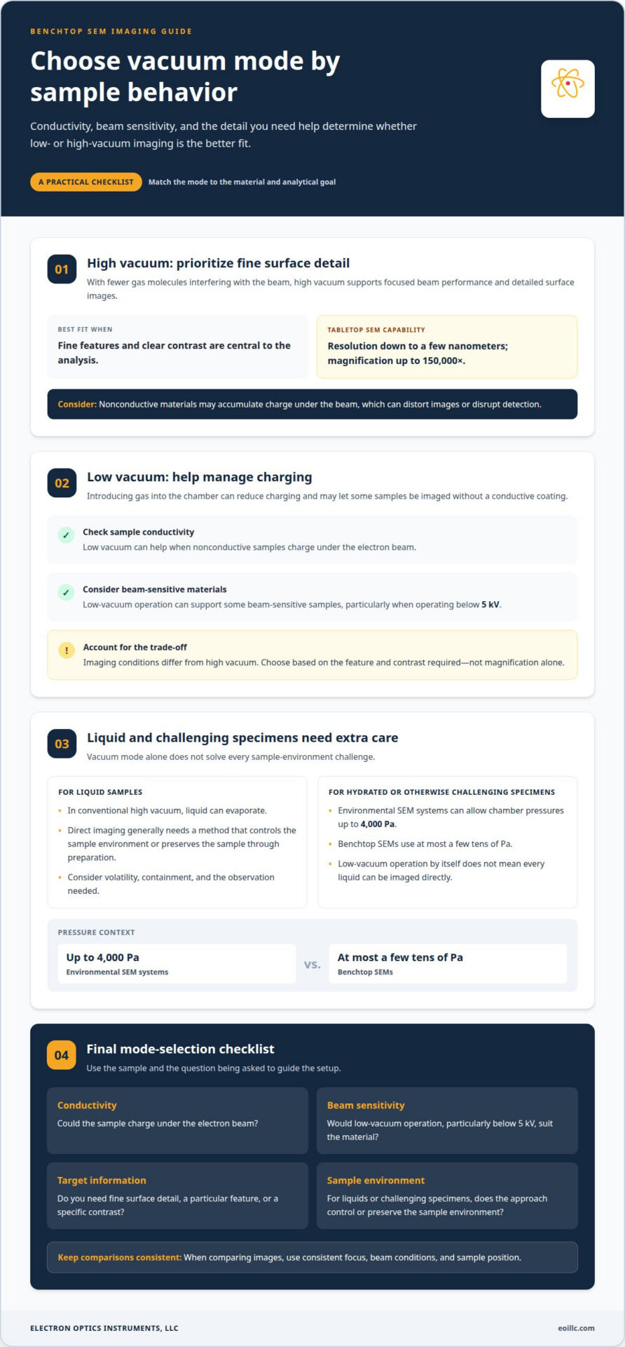
Vacuum mode affects how an electron beam interacts with a sample. In high vacuum, fewer gas molecules interfere with imaging, supporting focused beam performance and detailed surface images. Tabletop SEM systems typically provide resolution down to a few nanometers and magnification up to 150,000x. These capabilities suit applications where fine surface features and clear contrast are central to analysis.
Nonconductive materials can accumulate charge under the beam, distorting images or disrupting detection. Low-vacuum operation introduces gas into the chamber, helping reduce charging and allowing some nonconductive samples to be imaged without conductive coating. This mode can also support beam-sensitive materials, particularly when operating below 5 kV. The trade-off is that imaging conditions differ from high vacuum, so the best mode depends on the feature and contrast required.
Liquid samples need special consideration. In a conventional high-vacuum environment, liquid can evaporate, so direct imaging generally requires a method that controls the sample environment or preserves the sample through preparation. Low-vacuum operation alone does not mean every liquid can be imaged directly. Choose an approach based on the sample’s volatility, containment, and the observation you need.
Environmental SEM systems can allow chamber pressure levels up to 4000 Pa, while benchtop SEMs use at most a few tens of Pa. That difference matters for hydrated or otherwise challenging specimens. A university laboratory examining a polymer or biological material, for example, may choose low vacuum to limit charging, then use high vacuum for a conductive reference sample. Comparing images requires consistent focus, beam conditions, and sample position.
For routine tabletop imaging, choose the mode according to conductivity, beam sensitivity, and the information needed, not magnification alone. The Genesis Tabletop SEM is an option to consider for laboratory imaging workflows where tabletop SEM capabilities fit the analysis requirements.
Tabletop SEM accessibility and affordability
A tabletop scanning electron microscope brings electron microscopy into the working laboratory. Over the last two decades, benchtop SEMs have become more accessible to beginners, extending their use beyond specialist facilities. For example, undergraduate students at the University of California, Berkeley, use a new SEM in a teaching laboratory. This illustrates how an instrument can support research workflows and practical instruction when its capabilities match the questions being studied.
Accessibility also depends on workflow. Benchtop SEMs can allow companies to perform analyses in their own laboratories in as little as 15 minutes. That can help teams inspect a production sample during an investigation rather than wait for an outside analysis. The time required still depends on preparation, imaging conditions, and the information sought. A surface image may be a straightforward task, while elemental analysis requires suitable detector configuration and interpretation.
Assess affordability in the context of the full operation, not the purchase price alone. Consider the analyses required, detector options, sample throughput, staff training, facility needs, and ongoing maintenance. A laboratory focused on routine surface inspection, for example, may need a different configuration from a team seeking elemental information. Matching capabilities to actual work helps avoid paying for functions that do not support the application.
Choose an instrument configuration to suit the material and analytical objective. A microscope intended for routine imaging may not provide every capability needed for advanced characterization, and a benchtop format does not remove those technical limits. First define whether you need surface morphology, composition, or another form of materials analysis, then align the detector and workflow with that goal. This makes tabletop SEM technology more accessible in practice by connecting equipment selection to the laboratory’s real needs.
How have benchtop SEM automation and AI-assisted analysis advanced?
Automation can reduce repetitive setup steps in a benchtop SEM workflow. A configured system may guide an operator through choosing a sample location, setting imaging conditions, focusing, and capturing images. For example, a technician inspecting several regions on one component can follow a repeatable sequence instead of setting up each acquisition from scratch. Consistent settings make image comparisons more meaningful, while the operator remains responsible for choosing suitable conditions.
Automated stage movement can support systematic examination. An operator can define multiple points across a sample and acquire images at each location using a consistent approach. This can help when checking a coating at its center, edges, and visible defects. Automation cannot determine whether those locations represent the whole component, so the sampling plan must reflect the analytical question and the material’s variation.
AI-assisted analysis can help organize and interpret image data. For example, image-analysis software may segment visible particles, pores, or cracks and measure their area or distribution. This can make repeated comparisons more efficient than marking every feature manually. Results still depend on image quality and the software’s training or classification settings. A bright feature may reflect a topographic or compositional difference, so interpret software output in light of the sample and imaging conditions.
For reliable results, use a controlled sequence: prepare and mount the sample, select representative fields, capture images under consistent conditions, and review automated measurements against the original images. If you use elemental analysis, distinguish image-based classifications from EDS results, which provide different information. Keep acquisition settings and analysis parameters with each dataset so later comparisons use the same basis. Automation can improve repeatability, but it does not replace an operator’s judgment about artifacts, contrast, or meaningful features.
To discuss SEM systems and analytical workflows for your laboratory, contact Electron Optics Instruments, LLC.
Electron Optics Instruments, LLC: Cube II Benchtop SEM
Electron Optics Instruments, LLC offers the Cube II Benchtop SEM for laboratories considering benchtop scanning electron microscopy. It may suit your work when you need in-house materials imaging and are assessing whether a benchtop instrument fits your samples and analysis workflow.
Choose based on the analytical task. Consider your sample types, imaging requirements, and whether EDS elemental analysis is part of your workflow. Electron Optics Instruments, LLC also offers the Genesis Tabletop SEM, Veritas Series SEM, and Veritas Plus SEM, giving you additional SEM options to consider alongside the Cube II. Evaluate each system against your intended applications rather than assuming one suits every specimen or analytical need.
If you’re evaluating benchtop SEM options for materials analysis, contact Electron Optics Instruments, LLC to discuss your requirements.
Turn analytical goals into a practical SEM workflow
Start by defining what a successful analysis must deliver. Identify your sample types, the images or measurements you need, and how the results will inform a materials decision. This gives you a practical basis for matching instrument capabilities to laboratory needs, rather than choosing a system by magnification or automation alone. Advances in benchtop SEM technology matter most when they support a repeatable workflow and answer a specific analytical question.
Electron Optics Instruments, LLC offers the Cube II Benchtop SEM, Genesis Tabletop SEM, and Veritas Series SEM. These options provide a starting point for discussing how benchtop SEM equipment can suit your intended applications and analysis priorities. Begin with your materials, workflow, and the information you need.
Contact Electron Optics Instruments, LLC to discuss your SEM requirements and take the next step toward a more informed materials analysis workflow.
Frequently Asked Questions
What is a Tabletop SEM?
A tabletop SEM is a compact scanning electron microscope designed to fit on a laboratory bench. It scans a focused electron beam across a sample and detects the resulting signals to create detailed images. Some configurations also support analytical methods such as EDS, which can provide information about elemental composition. Capabilities depend on the system’s configuration and the material being examined.
What is the difference between a tabletop and full-size SEM?
A tabletop SEM has a compact format, while a full-size SEM generally provides a larger platform for instrument configurations and sample handling. The practical question is whether the system accommodates the specimens, detectors, and analytical tasks you need. A laboratory studying small prepared samples, for example, may prioritize a compact setup, while work with larger components may call for a system designed around those dimensions.
What magnification can a compact SEM achieve?
Magnification varies by instrument and configuration. The Cube II SEM is specified for magnification up to 300,000×, giving users a way to examine fine features across a range of scales. Magnification alone doesn’t establish the smallest feature that can be resolved: image quality also depends on resolution, focus, sample preparation, and operating conditions. Select magnification according to the detail the analysis needs to reveal.
Why Choose a Compact or Desktop SEM?
A compact SEM can suit laboratories that need electron microscopy within their workflow and have samples compatible with a benchtop instrument. It can support routine inspection, teaching, or materials investigations without requiring a full-size installation. For instance, a research group can examine prepared specimens during a project, then assess whether the system’s imaging and detector options address its specific analytical question.
What are the latest advances in benchtop SEM technology?
Advances in benchtop SEM technology include broader options for low-vacuum imaging, analytical detector integration, and automated image acquisition. Some systems support nonconductive sample observation, while EDS adds elemental analysis to electron imaging. Automation can help standardize repeat image capture, but scientists still need to select representative regions and interpret results. The key is matching imaging and analysis capabilities to the sample.
How have benchtop SEM resolution and magnification improved?
Current benchtop SEM specifications vary, and magnification and resolution describe different performance characteristics. Some benchtop systems are available with 5 nm resolution, while the Cube II SEM is specified at 5 nm resolution and up to 300,000× magnification. Higher magnification enlarges the displayed image; resolution determines how closely spaced features can remain distinct. Compare both measures with the features and sample conditions in your analysis.