
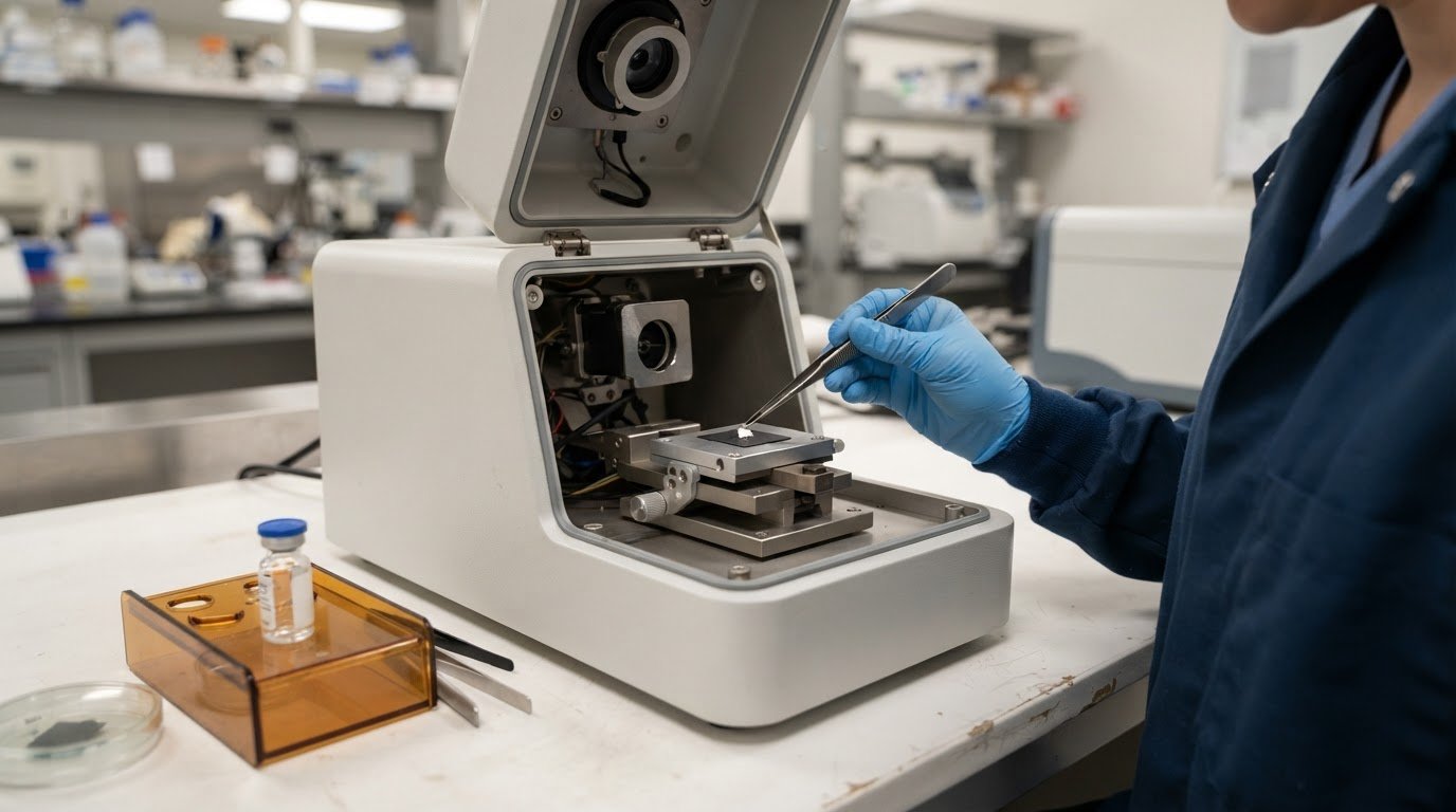
A product can appear clear while containing particles too small to see. USP <788> defines particulate matter in injections as mobile, undissolved particles, other than gas bubbles, that are unintentionally present in solutions. Identifying foreign particulate matter can reveal fibers, metal fragments, glass, polymers, or other contaminants, including particles as small as 20 μm.
Visual inspection is a useful first step, but it can miss particles: according to the United States Pharmacopeia, it detects fewer than 5% of particles smaller than 50 μm. You’ll learn how microscopy documents a particle’s size and shape, how spectroscopy helps determine its composition, and how those findings can guide an investigation into potential contamination sources. The article also explains how these complementary techniques support a more informed assessment of pharmaceutical particulates.
Key Takeaways
- Recognize the range of particles that can affect pharmaceutical products, from fibers and metal fragments to material formed through crystallization or degradation.
- Use microscopy to document particle size and morphology, then apply spectroscopy to help characterize its chemical composition.
- Consider equipment, packaging, raw materials, and the production environment when tracing a particle’s likely origin.
- Learn how SEM and energy-dispersive spectroscopy systems can support detailed particle examination and contamination investigations.
What are foreign particles in pharmaceutical products?
Foreign particles are unintended, undissolved material in a product, rather than part of its formulation. USP <788> defines particulate matter in injections as mobile, undissolved particles, other than gas bubbles; identification and characterization combine observation with methods that examine morphology and composition.
U.S. pharmacopeial standards address different contexts: USP <788> covers injections, USP <787> therapeutic protein injections, and USP <789> ophthalmic solutions. USP <790> covers visible particulates in injections, while USP <1788> covers methods for particulate matter determination. Visual inspection detects fewer than 5% of particles smaller than 50 μm, according to the United States Pharmacopeia.
Elemental and Crystalline Particle Analysis
For inorganic particles, scanning electron microscopy (SEM) with energy-dispersive X-ray spectroscopy (EDS) combines detailed imaging with elemental information. This can help distinguish, for example, a metal fragment from a glass shard. X-ray powder diffraction examines crystalline structure and can help characterize salts, polymorphs, or excipient-related contaminants. Choose a method based on the particle and the question the investigation needs to answer.
Small and Complex Particle Challenges
Particles may be as small as 20 μm, while manufacturing contaminants commonly range from 50-300 μm and can reach a few millimeters. Small size, mixed materials, and irregular morphology complicate isolation and analysis. A particle’s appearance alone rarely establishes its origin. Raman spectroscopy can provide a molecular fingerprint and is non-destructive for surface analysis, preserving material for further examination.
Collect the particle carefully in a clean environment, avoiding tools and surfaces that could introduce debris. If product residue needs to be removed, particle-free water may be used, provided the rinse does not compromise the sample. Record the particle’s appearance and location, and document how it was handled. Images and analytical findings make it easier to compare particles and assess possible sources.
Particle analysis can also inform manufacturing procedures and inspector training. If recurring morphology or composition suggests equipment wear, use the findings to guide equipment checks and process controls. For solvent-soluble particles, LC-HRMS may be considered as part of a broader investigation. Together, these approaches help distinguish visible defects from less apparent contamination and support manufacturing decisions.
Microscopy and spectroscopy for particle identification
Microscopy records a particle’s visible features; spectroscopy adds chemical evidence that can help characterize its composition and possible source. For identifying foreign particulate matter, laboratories may combine optical microscopy, SEM-EDS, FTIR microscopy, or Raman spectroscopy. Select methods according to the sample and the investigation’s objective.
For an unknown particle, document its appearance, then combine imaging with elemental or molecular analysis. SEM-EDS provides elemental information, while FTIR and Raman can help identify organic materials, including polymers.
Visual Particle Characterization
Optical or confocal microscopy can reveal particle size, shape, color, surface features, and distribution. Polarized microscopy shows how a particle interacts with polarized light, which can help distinguish visually similar materials. Photomicrographs and image analysis preserve observations for comparison across samples. These records help guide the choice of analytical techniques and show whether particles found during production share a pattern.
Particle Morphology Assessment
Morphology assessment examines whether a particle is fibrous, flake-like, spherical, or irregular, as well as its edges and surface texture. Twists and turns in a fiber, for example, may help distinguish natural cellulose from synthetic material, but appearance alone cannot establish identity. Comparing particle morphology can reveal similarities and guide chemical testing and root-cause investigation.
For chemical imaging, FT-IR microscopy collects infrared spectra from selected particle regions; spot analysis can be performed on particles above 0,1 mm. Raman spectroscopy can examine particles a few microns in size and provide a molecular fingerprint. Its probe size can be approximately 1 µm, enabling analysis of localized features. Raman surface analysis is non-destructive and requires no sample preparation.
Microplastics and other polymer fragments are organic materials, so FTIR or Raman can help distinguish their chemical signatures. SEM-EDS systems, by contrast, can characterize elemental composition and pair it with high-resolution images, supporting examination of inorganic matter. A workflow for identifying foreign particulate matter may use EDS systems when elemental evidence can help characterize a particle.
Potential sources of contamination
A particle’s material and morphology can help narrow its possible origin, but pharmaceutical investigations also examine where it may have entered the process. Raw materials, equipment, production environments, packaging, and transport each present distinct contamination pathways.
Equipment wear can release metal abrasion from pipes, pumps, or vessels; corrosion may produce iron-containing particles. Filters, joints, and gaskets can shed material, while glass vials, rubber stoppers, and plastic components are possible packaging-related sources. Dust or fibers may enter during transportation or storage. Raw-material impurities, cross-contamination, human error, and tampering are also possible sources, but appearance alone does not establish any of them.
Formulation and storage matter, too. Crystallization, precipitation, or degradation of an active ingredient or excipient can create unwanted particles without an external contaminant entering the product. For example, finding a particle formed during storage may direct the investigation toward product stability rather than equipment wear. Particles in parenteral or inhalation products merit particular attention because these routes can introduce material directly into the body.
A lifecycle assessment follows the product from incoming materials through manufacturing, packaging, shipping, and storage. Compare when a defect first appears with equipment inspections, maintenance, repairs, cleaning records, and packaging changes. Repeated metallic particles near a pump, for instance, make inspection of that process area relevant; fibers appearing after packaging focus attention elsewhere. These patterns guide source investigations, but particle identity and process evidence must be considered together.
Counting and size assessment depend on the product category and method. USP <788> addresses particulate matter in injections; USP <787> covers subvisible particles in therapeutic protein injections, and USP <789> covers ophthalmic solutions. USP <790> addresses visible particulates in injections, while USP <1788> covers determination methods. These standards provide different contexts for particle assessment, rather than one universal size threshold for every pharmaceutical product.
Visual inspection can flag affected units, while microscopy and analytical techniques support particle sizing and characterization. Tracking findings across batches can reveal recurring patterns, inform manufacturing procedures, and refine inspector training as particulate conditions change. Cleaning, environmental controls, and equipment inspections before and after production help address identified pathways; maintenance and operator training support those controls.
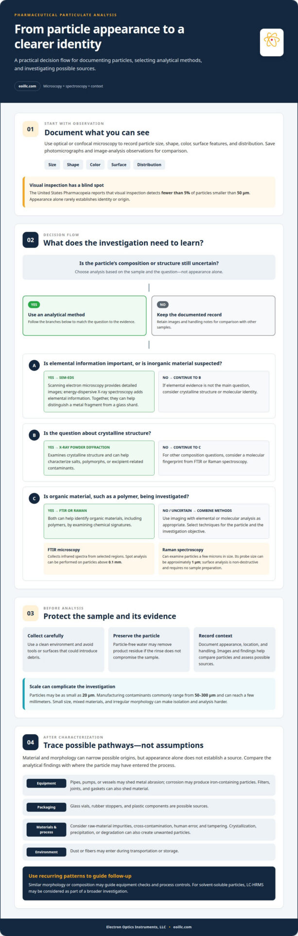
Identifying and tracing particulate contaminants
A sound investigation connects particle evidence with the product, process, and sampling context. The FDA expects manufacturers to investigate observed contamination, and regulatory evaluation also considers the product’s intended use and potential patient impact.
To distinguish an unwanted particle from an ingredient, compare its morphology and chemical signature with the formulation’s expected materials. Then examine environmental controls and appropriate background samples. A particle that matches a known excipient may reflect product composition; a different elemental or molecular profile may indicate extraneous material. Microscopy and spectroscopy provide complementary evidence, but do not determine a source on their own.
Begin with controlled sampling. Record where and when the particle was found, the affected unit or batch, and how the sample was handled. Preserve representative material and avoid contact with tools or surfaces that could introduce debris. Photomicrographs and image analysis document visible features, while elemental or molecular results strengthen characterization. Clear records make comparisons across samples more reliable.
Next, compare the finding with process evidence. Review relevant equipment, filter, joint, gasket, and packaging inspections alongside maintenance, cleaning, and production records. If similar particles appear near a process component, investigate that pathway; if they occur across unrelated lines, consider shared environmental or raw-material sources. A match in composition and morphology can guide follow-up, while timing and process evidence help assess the connection.
For injections, USP <790> addresses visible particulates, and USP <1788> covers determination methods. Visible findings should be tracked alongside analytical results, including particles from units rejected during inspection. This helps teams assess patterns, refine inspector training, and choose appropriate corrective action. The FDA and EMA expect manufacturers to investigate observed contamination; findings can inform risk assessment and product-quality decisions.
Where elemental evidence can narrow a suspected source, explore EDS systems for particle characterization. Results can help distinguish materials such as metal or glass, supporting a focused investigation of equipment, packaging, or environmental pathways.
Electron Optics Instruments
Electron Optics Instruments offers scanning electron microscopes for laboratories investigating pharmaceutical particles. When optical observations need higher-resolution imaging, SEM can help examine particle surface features and morphology as part of a broader identification workflow.
Options include the Cube II Benchtop SEM, Genesis Tabletop SEM, Veritas Series SEM, and Veritas Plus SEM. These systems provide SEM imaging to support examination of particle form and structure. Select an instrument according to the sample and the analytical questions the investigation needs to answer.
SEM imaging contributes evidence, but does not alone establish a particle’s chemical identity or contamination source. Consider it alongside other analytical methods and process records when building a complete investigation. Contact Electron Optics Instruments to discuss SEM options for pharmaceutical particle examination.
Turn Particle Findings Into Better Decisions
The next step is to connect laboratory evidence with decisions that strengthen quality systems. A well-documented investigation can help teams determine whether to collect additional samples, review a process more closely, or change inspection practices. Connecting analytical results with operational action is central to identifying foreign particulate matter effectively.
When evaluating SEM imaging for particle analysis, consider whether the instrument can address the questions raised by your samples and provide the detail your investigations require. Electron Optics Instruments offers SEM options for this work, including the Cube II Benchtop SEM, Genesis Tabletop SEM, and Veritas Series SEM.
Discuss your laboratory’s particle-analysis needs and SEM options directly with Electron Optics Instruments. A focused conversation can help clarify a practical next step for your analytical workflow.
Frequently Asked Questions
What is Foreign Matter?
Foreign matter is material in a product that is not intended to be part of its composition. It may enter from outside or arise when the product or one of its ingredients changes. The distinction matters: a particle resembling a formulation ingredient may still warrant investigation if its location, appearance, or distribution differs from expected product characteristics.
Where Do Contaminants Come From?
Contaminants can enter at different points in a product’s lifecycle or develop within the product during storage. Compare where the particle was observed with when the product was made, packaged, transported, and stored. Findings limited to one batch, for example, may point to a batch-specific process event, while a broader pattern may suggest a shared material or handling pathway.
What methods are used to identify foreign particles in food, pharmaceuticals, and other products?
Choose methods according to the sample and the identity question. Microscopy documents physical features; SEM-EDS combines imaging with elemental information, while FTIR or Raman can help characterize molecular composition. For soluble material, LC-HRMS may be considered; X-ray powder diffraction examines crystalline structure. In food, pharmaceutical, or device investigations, interpret analytical findings alongside product composition and process context.
How do visual inspection and microscopy compare for identifying particulate matter?
Visual inspection provides a direct way to screen products for observable defects, while microscopy magnifies and records particle features for closer examination. Microscopic images support comparisons between samples and help determine which analytical method to use next. For example, document a visible fiber’s shape and surface detail before chemical testing to help identify its material.
What are common sources of foreign particulate contamination in manufacturing?
Common source categories include incoming materials, process components, packaging, and the production environment. The practical task is to connect a particle’s characteristics with the manufacturing stage where it could have entered. Compare findings across affected units and batches, then review changes in equipment condition, materials, or handling. This evidence-based approach helps prioritize source checks without assuming that a single particle proves a specific failure.