COVID-19 has changed the way we live, work, and even the way we think about public health on the grandest possible scale. Independent researchers around the world have shared hundreds of thousands of SARS-CoV-2 genome sequences in an effort to better understand new variants of the disease and how to combat them. This has been described as a game changer by the World Health Organisation (WHO) – but the genomic sequence of coronaviruses remains something of a mystery. Scientists have gained a good understanding of its transmission but question marks remain over its mutability.
Many successes in the ongoing battle to contain and eradicate COVID-19 have been enabled by workhorse analytical instruments like the electron microscope. Scanning electron microscopy (SEM) played an important role in probing and ultimately identifying key morphological features of SARS-CoV-2. SEM microscopes remain an invaluable tool for microbiologists and virologists studying potential targets for drug treatments and therapeutics. Additionally, small-scale SEM microscopes may help laboratory technicians get back to work safely by enabling social distancing in the lab.
How SEM Microscopes Helped Identify COVID-19
Although the COVID-19 pandemic was initially mischaracterised as a local outbreak of viral pneumonia in Wuhan, China, the global scientific community was quick to make up for lost time by putting SARS-CoV-2 under the microscope. SEM microscopes had been used to probe the morphological structure of the initial SARS-CoV virus, thus it was the natural tool for investigating the surface characteristics of the novel coronavirus.
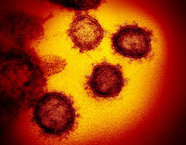
NIAID’s Rocky Mountain Laboratories (RML) in Hamilton, Montana, was one of the first facilities to publish images of SARS-CoV-2 that confirmed its similarity to other coronaviruses like MERS-CoV and SARS-CoV, with its characteristic surface structure of spike proteins.
Using SEM Microscopes for Therapeutic Development
Since the COVID-19 virus was first characterised, pharmaceutical and reagent manufacturers have accelerated their efforts to develop diagnostic and therapeutic solutions. SEM microscopes have again played a key role in rapid detection of SARS-CoV-2 while helping to uncover the replication cycle of the virus. Researchers in Marseille used SEM microscopy to follow the virus from 1 to 36 h post-infection, showing that the virus managed to infect healthy cells via membrane infusion, and were expelled from by either lysis or fusion.
How SEM Microscopes Help Labs Cope with the Virus
COVID-19 has not only impacted the scientific community by enforcing collaboration and round-the-clock investigations of a new viral strain, it has also imposed the same restrictions on labs as every other business. Mandatory social distancing and regular deep cleaning of workstations is as essential in laboratories as any other workplace. Although most labs have the benefit of strict, pre-established hygiene rules, most facilities equipped with large-scale SEMs will struggle to maintain pre-COVID workflows due to the new restrictions. Fortunately, small-scale SEM microscopes can help enforce social distancing in the lab.
If you would like to learn more about the strengths of SEM microscopes in combatting the spread of COVID-19, contact us today.
Further reading:
https://www.frontiersin.org/articles/10.3389/fmicb.2020.02014/full
https://www.niaid.nih.gov/news-events/novel-coronavirus-sarscov2-images
Image Credit: NIAID-RML