
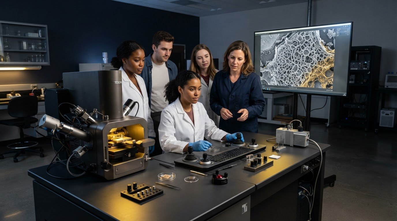
A striking microscope image does not, by itself, teach students how to interpret it. Introducing electron microscopy in university curriculum is more effective when students follow the whole process: posing a scientific question, preparing a sample, choosing imaging conditions, and evaluating what the resulting image can and cannot show. This approach gives students a foundation for using microscopy thoughtfully rather than treating the instrument as a black box.
Limited instrument access, specialist teaching time, and competing course demands can make that goal challenging. A successful introduction does not require every student to operate a microscope independently. It does require learning objectives that connect core concepts with meaningful practical exposure and assessment. This guide explains how to sequence the fundamentals, plan hands-on or shared-access learning, assess students’ understanding of image formation and limitations, and choose a course format and equipment strategy suited to institutional capacity. The result is a practical plan that balances theory, sample preparation, and imaging while keeping responsible instrument use in view.
Key Takeaways
- Teach electron microscopy as a connected workflow, linking research questions and sample preparation to image interpretation.
- Sequence SEM concepts so students move from electron-beam fundamentals and instrument components to image acquisition and evidence-based analysis.
- Choose demonstrations, supervised sessions, shared facilities, or digital resources according to course goals, cohort size, and available expertise.
- Make learning assessable by aligning each activity with observable evidence, such as an annotated image or a reasoned interpretation.
- Plan sustainable instrument access by comparing institutional purchasing, shared facilities, and external support against course needs, staffing, and maintenance responsibilities.
Why Introduce Electron Microscopy in the University Curriculum?
Introducing electron microscopy in university curriculum gives students a way to examine evidence at scales that visible-light microscopy cannot readily resolve. Instead of using visible light to form an image, an electron microscope uses a focused beam of electrons and detects signals produced through interactions between the beam and a specimen. The Electron microscope overview provides background on instrument types, including scanning electron microscopy (SEM) and transmission electron microscopy (TEM). In SEM, detected signals can reveal surface features and structural detail, but an image is not a direct or complete account of a specimen.
That distinction creates a useful teaching opportunity. Students can learn to describe what an image shows, consider how imaging conditions and sample preparation shape the result, and separate observation from interpretation. In materials science, they might examine fracture surfaces; in biology, compare specimen surface structures; in engineering, relate a component’s visible features to a design question. Interdisciplinary laboratory courses can develop the same reasoning skills across fields. The value comes from the question students investigate and the evidence they assess, not simply from having an instrument in the room.
Which student learning goals can electron microscopy support?
Set outcomes that students can demonstrate. For example, ask them to identify visible features, relate the scale bar to the dimensions under discussion, and support an interpretation with evidence from the image. Connect each task to a discipline-specific question and make clear that an SEM image has limits. Conceptual familiarity, such as explaining how an image is formed, is different from independently preparing samples or operating an instrument.
Where can microscopy fit within an existing course?
SEM can be introduced through an introductory laboratory, a materials course, research methods, or a capstone project. First map the prerequisites. Depending on the activity, students may need grounding in imaging, relevant material properties, or laboratory safety. An instructor can pilot one lesson using an image-analysis exercise or demonstration, assess whether students meet the intended outcomes, and use that evidence to guide any wider course change.
A measured introduction also helps match teaching goals to available expertise and access. If students are expected to operate equipment, plan for appropriate supervision and preparation. If the goal is image interpretation, an instructor-led demonstration or shared-facility experience may be sufficient. Define the learning objective before selecting the format, so instrument access supports the curriculum rather than becoming its purpose.
Build a Teaching Sequence Around How Scanning Electron Microscopy Works
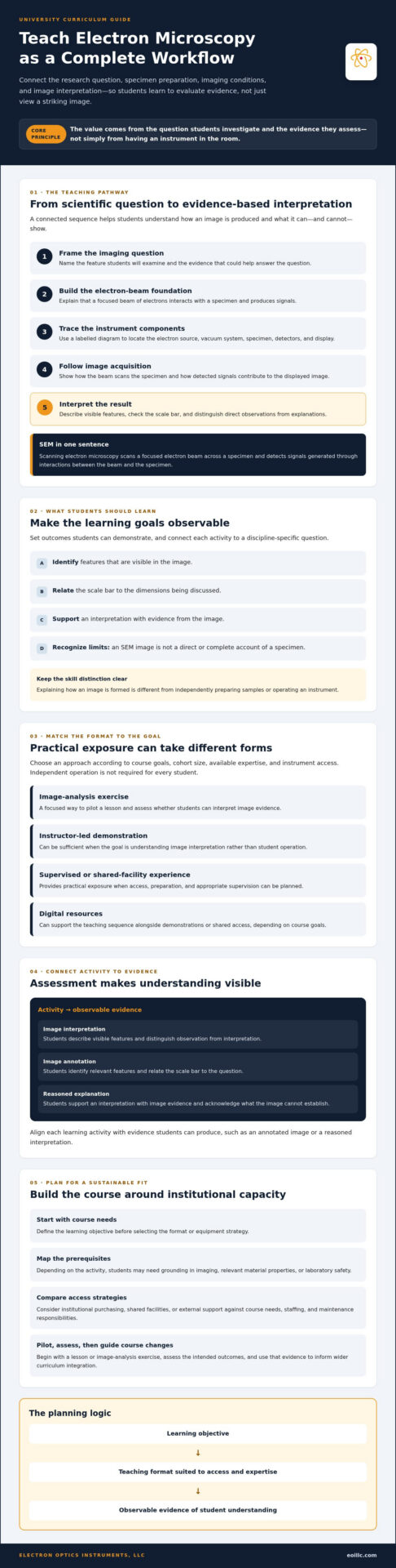
A clear teaching sequence follows the path from a scientific question to an interpretable image. Scanning electron microscopy (SEM) forms images by scanning a focused electron beam across a specimen and detecting signals generated through interactions between the beam and the specimen. An electron beam is a stream of electrons directed and focused onto a specimen inside the microscope. These definitions give students an accessible starting point before they encounter more detailed terminology.
Organize the lesson in five stages:
- Frame the imaging question: What feature should students examine, and what evidence would address the question?
- Introduce electron-beam basics: Explain how a focused beam interacts with the specimen to produce signals.
- Identify instrument components: Use a labelled diagram to trace the electron source, vacuum system, specimen, detectors, and display.
- Follow image acquisition: Show how the beam scans across the specimen and how the detector signal contributes to the displayed image.
- Interpret the result: Ask students to describe visible features, check the scale bar, and distinguish observation from explanation.
A diagram or labelled instrument image can make the sequence concrete. Have students trace the beam’s route and identify where signals are collected and converted into an image. Match the technical depth to the course level and stated outcomes. An introductory laboratory may need a functional account of the components, while an advanced course can examine imaging variables in greater detail. In either case, students should use magnification and scale bars carefully: magnification alone does not establish the actual size of a feature, and image detail needs to be interpreted in context.
Teach the SEM workflow before introducing advanced terminology
Begin with the specimen and question, then work through the process that makes an image possible. Introduce the source, vacuum system, detectors, and scanning process at the level students need to explain how a signal becomes an image. Ask students to annotate a schematic or image, connecting each component to its role instead of memorizing labels in isolation. This sequence makes technical language serve understanding.
Connect image interpretation to analytical methods
Students should first separate what they observe from what they infer. For example, they can describe a visible surface feature, then state what additional evidence would be needed to explain its composition or origin. EDS, or energy dispersive spectroscopy, can be introduced as an optional analytical extension for investigating elemental composition. It complements imaging fundamentals rather than replacing them. For a university-level example of structured microscopy instruction, Purdue outlines a practical introduction to the operation of transmission electron microscopes. Its focus is TEM, but its course framing can inform lesson planning.
When developing a practical SEM sequence, consider how equipment, technical training, and ongoing support fit the institution’s teaching objectives. Explore SEM equipment and support options from Electron Optics Instruments, LLC as part of that planning.
Choose a Teaching Format That Fits Instrument Access and Course Goals
The right format depends on what students need to demonstrate, not on whether every student can operate an SEM. A demonstration may support observation and interpretation; supervised practice is more appropriate when operating steps are an explicit learning outcome. Shared facilities and digital materials can extend access when local capacity is limited. Compare the options against cohort size, available teaching time, instrument access, and staff expertise before scheduling the activity.
| Format | Learning value | Access needs | Limitations | Assessment opportunities |
|---|---|---|---|---|
| Instructor-led demonstration | Observe workflow and connect settings to images | SEM and knowledgeable instructor | Students may have limited direct control | Annotated image, observation notes, questions |
| Supervised student session | Practise selected operating steps | Instrument, preparation time, and close supervision | Scheduling and staff capacity can constrain participation | Task checklist and justified imaging choices |
| Shared facility | See specialist workflows and facility practice | Coordinated access and approval | Procedures, availability, and allowable activities vary | Facility observation log and image interpretation |
| Digital resources | Review images and concepts flexibly | Suitable digital materials | Does not provide physical instrument experience | Image analysis and short written explanations |
What can students learn from a demonstration or shared facility?
Make observation active. Ask students to record the imaging question, the settings presented by the instructor, visible image features, and questions they would investigate next. A shared facility can provide access to instrument workflows when a department lacks teaching capacity or an SEM, but it is not automatically available for every course. Before describing a facility as an option, verify its procedures, scheduling, supervision, and access requirements with the institution.
When does hands-on SEM instruction make sense?
Use supervised practice when students are expected to gain operational familiarity. Define which steps they should perform rather than implying full independent operation. Allow for sample preparation, scheduling, instructor expertise, and equipment availability. A single session can introduce controls and workflow, but should not be presented as proof of independent proficiency. Institutions assessing equipment and training needs as part of introducing electron microscopy in university curriculum can review SEM equipment and training considerations.
Digital resources can prepare students for a demonstration or reinforce concepts afterward. They can also support image interpretation when instrument time is unavailable. Choose the format that produces assessable evidence for the stated objective, then adjust the activity to fit actual staffing and access.
Create a Practical Lesson Plan, Assessment, and Safety Framework
A reliable SEM lesson connects its learning outcomes, activity, assessment, and safety planning before students enter the laboratory. This sequence gives instructors a reusable structure for introducing electron microscopy in university curriculum while keeping each practical activity aligned with what students are expected to learn.
- Define outcomes: Specify what students should explain, identify, or justify by the end of the lesson.
- Select an activity: Choose a demonstration, guided image analysis, or supervised instrument task that directly supports those outcomes.
- Prepare materials: Assemble the image, prompts, sample, and assessment criteria. Confirm sample suitability and preparation requirements against institutional procedures and instrument guidance.
- Teach and assess: Brief students on the task, guide their observation, and collect evidence of learning.
- Review and revise: Use student work and instructor observations to identify unclear concepts, timing issues, or access constraints before repeating the lesson.
What should an introductory SEM lesson include?
Use a focused question, a short concept briefing, a guided observation, and a reflection. For example, students might examine an instructor-selected image and describe which visible features are relevant to a stated materials or biology question. They should explain what the image supports and what it cannot establish. If the activity includes physical sample preparation or instrument use, have qualified local staff review the procedure, sample constraints, and safety requirements under institutional and manufacturer guidance.
Before teaching, confirm that operating procedures, permitted samples, and safety instructions match local requirements. Faculty should involve the institution’s relevant safety personnel and SEM specialists when reviewing hands-on activities. A general lesson plan cannot replace equipment-specific procedures or local expert direction.
How can instructors assess student understanding?
Collect observable evidence, such as a labelled image, concise observation notes, and a short explanation grounded in image details. A simple rubric can distinguish three dimensions:
- Terminology: Does the student use key imaging terms accurately?
- Image reading: Can the student describe visible features and use the scale information appropriately?
- Reasoning: Does the interpretation follow from the evidence, with appropriate limits?
Reward careful uncertainty, not confident overclaiming. Ask students to identify possible artifacts, ambiguities, or unanswered questions, and distinguish these from direct observations. This makes responsible interpretation part of the assessed skill rather than an afterthought. For help planning instrument-based instruction, discuss SEM training and support options with Electron Optics Instruments, LLC.
Plan Sustainable Curriculum Integration and Instrument Support
A pilot lesson can establish whether SEM belongs in a course. Sustaining it requires clear ownership and realistic plans for instrument access, staff preparation, and ongoing support. Assign responsibility for coordinating the curriculum, preparing students, arranging facility access, and reviewing the lesson. Make these roles explicit so the activity does not depend on one instructor’s availability or informal knowledge.
Before considering equipment, document the intended sample types, student activities, learning outcomes, and expected use. Then compare possible approaches against those requirements:
- Shared institutional access may suit courses focused on image interpretation or occasional demonstrations, provided the facility’s procedures and scheduling can accommodate the class.
- Purchasing an SEM may be worth evaluating when repeated instrument access is central to course objectives and the institution can plan for installation, training, maintenance, and technical support.
- External support can be considered when the department needs equipment or training guidance. Confirm the scope, responsibilities, and availability directly.
How should a department evaluate equipment and support needs?
Compare capabilities with course requirements rather than selecting by model name alone. Consider whether planned activities call for demonstrations, supervised operation, or particular analytical capabilities. Verify instrument suitability against sample needs and current product details. Benchtop systems are one category to evaluate, not a universal solution; institutions can review desktop SEM capabilities alongside other access approaches. Include installation, user training, preventative maintenance, and technical support in the institution’s planning, and confirm responsibilities with relevant providers and campus staff.
How can a pilot course grow into a sustainable program?
Start with one course module and assess both student work and instructor observations. Gather feedback from faculty, technical staff, and participating students. Were the objectives achievable? Did access and preparation fit the course? Which concepts or procedures need revision? Use those findings to refine the module before extending it to other courses, and schedule curriculum review so activities keep pace with course needs and available support.
Training and service are institutional planning considerations, not guaranteed outcomes of purchasing a particular instrument. Electron Optics Instruments, LLC distributes SEMs and provides on-site technical training for laboratory staff. Departments evaluating equipment or training needs can contact Electron Optics Instruments, LLC for SEM information and confirm which options align with their requirements.
Turn a Pilot Lesson into a Sustainable Program
A durable SEM curriculum starts with a clear teaching purpose, then aligns the sequence, learning format, assessment, and safety review with that purpose. Students can build meaningful understanding through image interpretation and guided observation, even when independent instrument operation is not an objective. As the program develops, use student work and feedback from instructors and technical staff to refine the lesson and decide whether broader access is justified.
For departments considering equipment or training, compare options against actual course requirements and plan for ongoing responsibilities, not just initial access. Electron Optics Instruments, LLC offers SEM equipment, on-site technical training, and service support. These are factors institutions may wish to consider as they evaluate instrument-based instruction.
Contact Electron Optics Instruments, LLC to discuss SEM equipment and training as you plan the next step in introducing electron microscopy in university curriculum. A carefully scoped pilot can give your department a practical foundation for building instruction that is rigorous, assessable, and responsive to available resources.
Frequently Asked Questions
What is the best way to introduce electron microscopy to university students?
Start with a specific learning outcome, such as interpreting a surface image, and teach only the SEM concepts students need to achieve it. Pair a concise explanation with guided image observation and reflection. If instrument access is available, add a supervised demonstration or practice activity that fits the course objectives. Assess whether students can distinguish visible evidence from their interpretation, and ask them to identify what the image alone cannot establish.
Can electron microscopy be taught without an SEM on campus?
Yes. Labelled instrument diagrams, example images, recorded demonstrations, and access to a shared facility can introduce core concepts without a campus SEM. Use these resources to help students interpret evidence, discuss image limitations, and connect observations to a course question. They can support conceptual learning, but they do not provide direct operating experience. If independent instrument use is a learning outcome, arrange supervised access and assess that skill separately.
How should SEM be integrated into a university course?
Begin by identifying which existing course outcomes electron microscopy can support, then define the level of knowledge or skill students should demonstrate. Build a sequence from the imaging question through electron-beam concepts to image interpretation. Select an activity that fits available access and supervision, and assess student work against the intended outcomes. Review evidence from a pilot module before expanding it into a recurring laboratory or broader curriculum component.
What subjects can include electron microscopy?
Electron microscopy can support courses in materials science, biology, engineering, and laboratory research methods when image-based evidence is relevant to the subject. For example, students might examine material surfaces or biological structures, then relate their observations to a defined course question. Adapt the SEM theory and practical depth to students’ preparation and course level. Suitability depends on learning objectives and teaching capacity, not on a single activity being appropriate for every discipline.
Do students need hands-on SEM experience to learn electron microscopy?
No. Students can learn to interpret SEM images and explain the basic workflow through structured demonstrations, diagrams, and image-analysis exercises. Hands-on access is necessary when instrument operation is itself an intended learning outcome. In that case, plan for supervision, sample preparation, scheduling, and instrument availability. Set realistic expectations: a limited practice session can introduce operating steps, but should not be treated as evidence of independent proficiency.
How can instructors assess learning in an electron microscopy module?
Assess both what students observe and how they reason from the image. An annotated image, concise observation notes, or a short evidence-based interpretation can show whether they recognize scale, describe visible features, and support conclusions appropriately. Use a rubric that distinguishes terminology from image reading and reasoning. Include a prompt asking students to identify uncertainty, possible artifacts, or unanswered questions, so their conclusions remain within the evidence.
What should a university consider before buying an SEM for teaching?
Start with course outcomes, anticipated users, sample requirements, staff expertise, and the access time students will need. Evaluate instrument capabilities alongside installation, user training, preventative maintenance, and technical support. Compare options against the planned curriculum rather than assuming a particular model fits every institution. Electron Optics Instruments, LLC distributes SEM equipment and provides on-site technical training and service support. Verify current specifications and operating requirements with the manufacturer or distributor before procurement.