
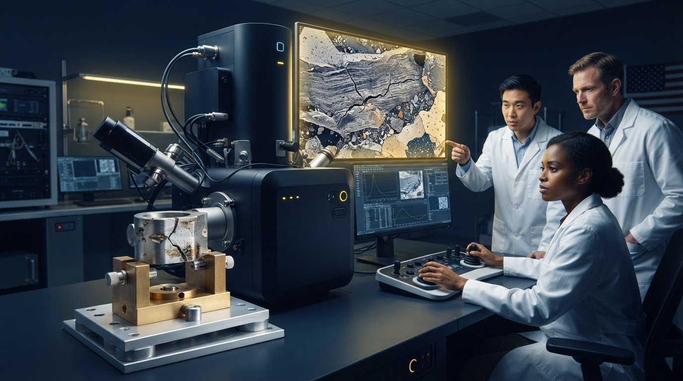
A visible defect is a starting point, not an explanation. A crack, residue, or unexpected surface feature can have several possible causes, leaving production teams without enough evidence to make a confident process change. Understanding how SEM helped solve a manufacturing problem starts with examining the defect at a scale that can reveal details conventional inspection may miss.
Scanning electron microscopy (SEM) produces detailed images of a sample’s surface, helping investigators compare defect features and narrow possible explanations. When composition matters, Energy Dispersive Spectroscopy (EDS) can add elemental information. Neither result establishes root cause by itself. Teams need to consider microscopy findings alongside process records, material history, and other relevant evidence.
This article explains how SEM imaging and EDS can support a manufacturing investigation, and how to connect those findings with production data. It also outlines a repeatable workflow for documenting observations, choosing corrective action, and checking whether the action addressed the suspected cause. The goal is to turn an unexplained defect into evidence that supports a more informed production decision.
Key Takeaways
- SEM imaging can reveal defect features that help turn an uncertain production issue into a focused investigation.
- SEM images and EDS elemental information can help narrow possible causes when composition is relevant.
- Understanding how sem helped solve a manufacturing problem means treating microscopy as one source of evidence, not proof of root cause by itself.
- Compare microscopy findings with sample history, process records, and other relevant tests before choosing corrective action.
- Document the investigation consistently, then verify whether the corrective action addresses the suspected cause.
How SEM Helps Turn a Manufacturing Defect into an Investigable Problem
A defect can be easy to see and difficult to explain. A recurring fracture, surface mark, or contaminant may have several possible origins. Changing a process before distinguishing among them risks addressing a symptom rather than its source. Scanning electron microscopy (SEM) helps make the defect investigable by producing detailed images of surface features that ordinary visual inspection cannot resolve.
SEM scans a sample with an electron beam to reveal surface morphology at high magnification. Its images characterize observable features, but they do not, on their own, confirm the production root cause. For an accessible overview of the instrument and its signals, see Scanning Electron Microscope (SEM). A clear understanding of how sem helped solve a manufacturing problem starts with this distinction: imaging provides evidence to test, not a verdict.
Which manufacturing questions can SEM help answer?
SEM can help teams examine whether a fracture surface has distinct regions or patterns, whether a particle has a particular shape or texture, and whether a coating appears uneven, cracked, or separated from its substrate. It can also reveal the size, distribution, and surface appearance of inclusions, scratches, and pits. These observations help classify what is present and guide the next investigation step. No single feature should be treated as a diagnosis without supporting evidence.
For example, if a production part shows a recurring surface mark, SEM imaging could help characterize its edges and texture. The team could compare affected areas with an unaffected reference, then use the differences to decide which possible causes to investigate. The comparison guides the inquiry without assuming what created the mark.
What SEM evidence can, and cannot, establish
An SEM image records morphology, allowing investigators to document and compare features across samples. Consistent sample selection and imaging conditions make those comparisons more useful, especially when assessing whether a defect looks similar across affected parts or differs from a reference sample.
Images alone generally cannot establish when a feature formed, which production step created it, or whether it caused a part’s failure. Interpretation depends on the sample’s history, process records, material information, and, where appropriate, complementary analysis. For foundational background on beam interactions and image formation, consult the article on scanning electron microscopy principles. The next step is to connect observed features to testable hypotheses, then compare those hypotheses with independent evidence before selecting corrective action.
How SEM Imaging and EDS Build a Stronger Defect Investigation
Once a defect has been defined, the next task is to collect evidence that can distinguish among plausible explanations. A disciplined SEM investigation moves from a specific question to representative samples, appropriate imaging, structured comparison, and interpretation. The question might be whether a particle differs from the surrounding material, whether a coating has separated, or whether fracture features vary between affected and unaffected parts.
From sample selection to interpretable images
Sample choice shapes what the team can conclude. Where available, compare affected parts with unaffected references and samples from relevant production stages. These comparisons can show which features are associated with the defect and when differences may appear. Preparation and imaging conditions can also affect what is visible, so document observations consistently and interpret them in light of how each sample was handled.
SEM images can be formed from different electron signals. Secondary-electron imaging typically emphasizes surface topography, making edges, texture, and fine surface features easier to examine. Backscattered-electron imaging provides contrast related in part to differences in average atomic number. This can help distinguish regions with different compositions, but the contrast is a clue for further investigation, not a standalone material identification.
When EDS adds useful compositional evidence
Energy Dispersive Spectroscopy (EDS) complements SEM imaging by detecting characteristic X-rays and indicating which elements are present in an analyzed area. This can help assess whether a particle’s elemental profile differs from the substrate, whether a suspect region contains elements associated with contamination, or whether two material areas appear compositionally distinct. For more on advanced SEM analytical techniques, consider which complementary methods can address questions beyond surface appearance.
EDS findings still need context. Elemental information may help narrow hypotheses, but it does not necessarily identify a unique source or explain how material arrived at a defect. Sample composition, location, comparison data, and process history all matter.
SEM observations support, but do not independently prove, the root cause of a manufacturing defect. The practical value of understanding how sem helped solve a manufacturing problem lies in combining image evidence with compositional findings, sample comparisons, and production records, then testing whether the interpretation fits the full evidence. This keeps a striking image from becoming an unsupported conclusion and gives the team a stronger basis for deciding what to investigate next.
Electron Optics Instruments, LLC offers SEM and EDS systems for manufacturing investigations. Explore the options at SEM and EDS systems.
Can SEM Identify the Root Cause Alone? Compare Evidence Before Deciding
No. SEM can narrow hypotheses by showing defect features at high magnification, but a micrograph alone rarely establishes the complete production cause. An image may reveal a particle, crack, or coating separation. Determining how and when it formed requires comparison with other evidence. This distinction helps teams use microscopy to guide decisions without treating a persuasive image as proof.
Make each analysis step answer a defined question. Use SEM to characterize fine morphology, EDS when elemental composition could distinguish hypotheses, and optical inspection to assess broader visible patterns. Process records can show whether a feature aligns with a material lot, production stage, equipment condition, or operating change. Use other tests when a specific uncertainty remains and the result could affect the next decision.
Separate observation from interpretation
- Observed evidence: A documented image shows a particle at a fracture edge, or a coating discontinuity at a sampled location.
- Plausible interpretation: The feature may relate to contamination, material separation, or a process condition. These are hypotheses to evaluate, not confirmed explanations.
- Confirmed cause: A conclusion supported by representative sample comparisons and independent process evidence that fits the suspected mechanism.
Match evidence to the unresolved question
Before adding another analysis, identify what the team still needs to know. If the uncertainty concerns the shape or distribution of a fine surface feature, SEM may be appropriate. If elemental differences could separate competing explanations, EDS may add useful information. If the question concerns when the issue began, review production timing, material records, and equipment conditions. Additional testing is justified when it can resolve an important uncertainty, not simply because another technique is available.
False certainty can arise from a nonrepresentative sample, preparation artifacts, or a feature found at only one isolated location. Compare affected parts with suitable controls and ask whether the observation recurs across relevant samples. A striking image is a starting point. A repeatable pattern, interpreted alongside process history, is stronger evidence.
In practical terms, how sem helped solve a manufacturing problem depends on connecting each observation to a decision: what to compare, which hypothesis to test, or whether a corrective action needs verification. Record what was seen separately from what it might mean, and describe a mechanism as confirmed only when independent evidence supports it.
A Practical SEM Workflow for Tracing a Manufacturing Defect
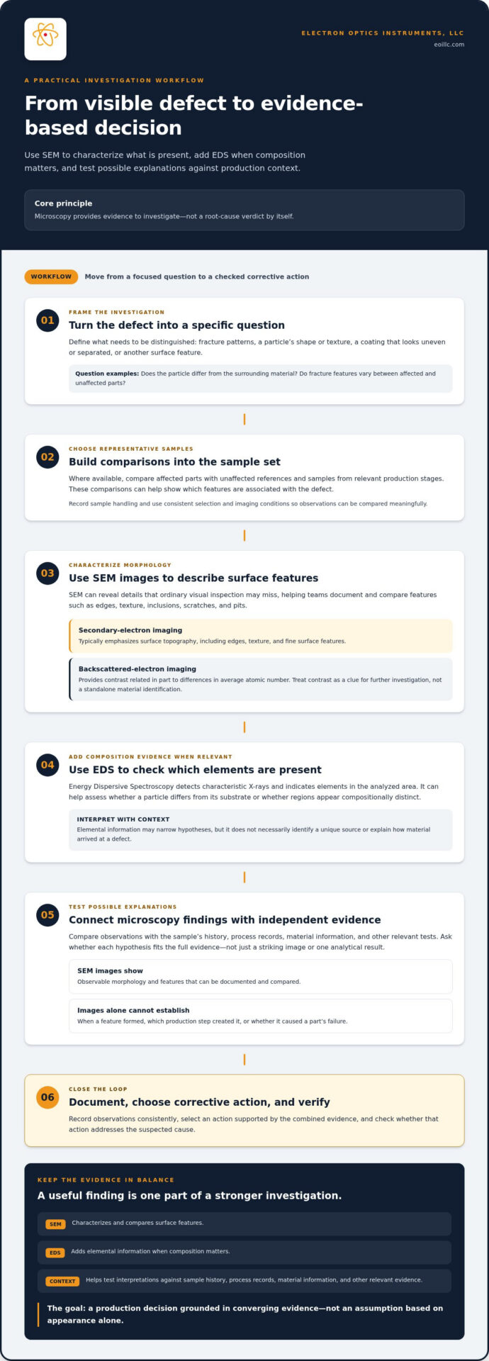
A repeatable workflow helps quality, engineering, and microscopy staff move from a production symptom to a verified decision. It also creates a record that distinguishes what the team observed from what it inferred. Use the steps below as a practical structure, adapting sample handling and imaging procedures to the material and investigation.
- Define the defect and its impact. Record where it occurs, how often it is found, when it first appeared, and which materials or production conditions are involved. Note relevant changes in equipment, inputs, or operating conditions.
- State the question and hypotheses. Before imaging, write down what the team needs to learn and which explanations it intends to assess. This helps keep interpretation tied to evidence rather than shaped after seeing a compelling image.
- Select and label representative samples. Preserve affected samples alongside appropriate unaffected or process-stage comparisons. Use traceable identifiers and record material identity, collection point, handling, and preparation so observations can be related back to production history.
- Image and document observations. Record sample identity, preparation, relevant imaging conditions, and observed features. Quality and engineering teams can then compare findings with the defect definition and original hypotheses. For guidance on consistent instrument use, consult SEM operation best practices.
- Apply a decision gate. If SEM evidence leaves competing explanations, identify the specific uncertainty before adding analysis. EDS may be relevant if elemental information could distinguish hypotheses. Process records or another test may be more useful if the unresolved question concerns timing or operating conditions.
- Choose, then verify, corrective action. Compare the findings with the process history and select an action that addresses the best-supported mechanism. Before implementing it, define a measurable follow-up, such as tracking defect frequency or applying a consistent inspection criterion to subsequent production.
Root-cause confidence is strongest when microscopy observations converge with representative sample comparisons and independent process evidence. After the change, review the agreed measure against the baseline and document whether results support the expected improvement. If they do not, reopen the investigation rather than treating the initial explanation as settled.
This is how SEM helped solve a manufacturing problem in a disciplined investigation: each observation informs a next decision, while verification tests whether the corrective action is effective. Electron Optics Instruments, LLC offers SEM systems and related support for investigative workflows. Explore SEM systems and support.
Apply SEM Findings to Production Decisions with the Right Instrument Support
Microscopy findings are most useful when teams can examine samples consistently and carry conclusions into production decisions. Access to an SEM, trained operators, and planned maintenance can help establish a dependable investigation workflow, from sample handling and image documentation to follow-up comparisons. Instrument support does not replace sound analysis, but it can help teams apply procedures consistently over time.
Assess whether benchtop SEM fits the investigation
Instrument suitability depends on the sample, the question under investigation, and the evidence the team needs to collect. Consider sample dimensions, the features of interest, whether elemental information could help distinguish explanations, and how examination will fit into the quality workflow. A benchtop SEM may support in-house examination when its capabilities align with those requirements. Base the decision on the investigation, not on a general assumption about the instrument category.
The Cube II Benchtop SEM is one of the SEM options offered by Electron Optics Instruments, LLC. For teams evaluating desktop systems, understanding desktop SEM capabilities and applications can help frame questions to consider during instrument selection.
Build a reliable workflow around instrument and expertise
Consistent operation depends on more than access to equipment. Staff training can help standardize sample handling, instrument use, and image documentation, making observations easier to compare across operators and investigations. Electron Optics Instruments provides SEM training, while preventative maintenance and repair support help maintain instrument operation as part of an ongoing microscopy workflow.
Where elemental evidence is relevant, EDS can complement SEM imaging with compositional information. Electron Optics Instruments offers EDS systems alongside benchtop and tabletop SEMs, supporting teams in aligning instrumentation with the analytical questions they need to address.
In practice, how sem helped solve a manufacturing problem is not only a question of image quality. It also involves selecting an instrument suited to the samples and questions, establishing consistent procedures, and preserving the ability to investigate and verify findings over time. Electron Optics Instruments, LLC offers SEM sales, EDS options, training, preventative maintenance, and repairs to support these workflows.
Explore SEM solutions and support for your manufacturing investigation workflow.
Turn Microscopy Evidence into More Confident Production Decisions
SEM can make an unexplained defect easier to investigate by revealing fine surface features and supporting comparisons between affected and reference samples. EDS can add elemental information when composition may help distinguish possible explanations. Neither replaces process records or verification. Microscopy findings are more useful when interpreted alongside production evidence and followed by a check that the corrective action addressed the suspected cause.
That is the practical value behind how sem helped solve a manufacturing problem: not a single image delivering an instant answer, but a structured path from observation to evidence-based action. Reliable instrument operation, consistent procedures, and trained staff help support that process.
Electron Optics Instruments brings more than 30 years of industry experience and is the sole US distributor for EmCraft Scanning Electron Microscopes. Its support includes SEM service, preventative maintenance, repairs, and on-site training, helping organizations maintain an effective microscopy workflow.
Explore the instrumentation and support options for your investigation. Explore SEM solutions and support, and take the next step toward clearer evidence and more informed production decisions.
Frequently Asked Questions
How can SEM help solve a manufacturing problem?
SEM helps solve a manufacturing problem by revealing fine surface features that clarify what a defect looks like and guide the next investigative step. Images may help characterize a fracture, particle, coating separation, or surface damage. Understanding how sem helped solve a manufacturing problem also means recognizing that the image is one part of the evidence. Compare it with reference samples, material information, and production records before deciding on corrective action.
Can SEM identify the root cause of a defect by itself?
No. SEM images alone rarely establish the complete root cause of a manufacturing defect. They show morphology and can support comparisons between affected and unaffected samples, but they do not necessarily reveal when a feature formed or which process step produced it. EDS may add elemental information when relevant. A defensible conclusion combines microscopy findings with process history, sample comparisons, and other evidence that supports the proposed cause.
What manufacturing problems can SEM investigate?
SEM can help investigate defects involving fracture surfaces, particles, coatings, inclusions, and surface damage such as scratches or pits. It can reveal the shape, texture, distribution, and fine details of these features, helping teams classify what they observe. Those observations can guide further analysis, but they do not prove a particular mechanism by themselves. Relate sample findings to the material, process stage, and production conditions.
How does EDS support SEM failure analysis?
EDS complements SEM imaging by detecting characteristic X-rays from a sample and indicating which elements are present in the analyzed area. This information can help assess whether a particle differs compositionally from the surrounding material or whether two regions have different elemental profiles. EDS results need context, however, and may not identify a unique source on their own. Compare them with sample location, material information, and process records.
What happens if SEM images show a defect but not its cause?
Treat the image as evidence that narrows the investigation, not as a final diagnosis. First, check whether the feature appears in representative affected samples and whether it is absent or different in appropriate controls. Then revisit the original hypotheses and identify what evidence would distinguish them. Depending on the unresolved question, EDS, process records, or another targeted test may help. Record observations separately from interpretations to avoid overstating certainty.
Is a benchtop SEM suitable for manufacturing quality investigations?
A benchtop SEM can be suitable when its capabilities fit the sample dimensions, analytical question, and evidence required for the investigation. Consider whether the work calls for fine surface imaging, complementary elemental analysis, or repeated in-house examination, and assess how the instrument would fit the team’s workflow. Electron Optics Instruments offers the Cube II Benchtop SEM. Suitability depends on the specific application, not the instrument category alone.
How can a manufacturer verify that an SEM-informed corrective action worked?
Before making a process change, define a measurable follow-up that relates directly to the defect, such as tracking its frequency or applying a consistent inspection criterion to subsequent production. Compare results with a documented baseline over an appropriate production period, while recording relevant process conditions and sample observations. If the defect persists or the evidence does not match expectations, revisit the suspected cause. Verification helps distinguish an effective correction from an unsupported assumption.