
What can a peer-reviewed paper using a benchtop SEM actually prove about the instrument? It shows how a system was used to investigate a particular sample and research question under reported conditions. How useful that evidence is for another laboratory depends on the instrument, operating conditions, sample preparation, and analysis described. Abstracts often leave out details that matter when judging whether a published method fits a different material or research goal.
Broad SEM searches can make benchtop studies difficult to identify, and an application demonstrated in one paper is not a universal measure of system performance. This article explains how to find relevant research, evaluate its methods and limitations, and compare study requirements with laboratory needs. It also offers a practical framework for assessing the Cube II Benchtop SEM against your requirements, without suggesting that it was used in the studies discussed.
Key Takeaways
- Combine “benchtop,” “tabletop,” or “desktop SEM” with a sample, method, or research question to find relevant studies.
- Judge a paper’s relevance by comparing its sample, instrument configuration, and methods with your research needs.
- Read the full paper for imaging settings, sample preparation, analytical methods, results, and limitations before applying its findings.
- Separate observations from SEM images from results produced by EDS or other analytical techniques.
- Translate relevant published methods into a laboratory requirements list covering samples, imaging goals, analytical needs, throughput, and users.
What Peer-Reviewed Papers Using Benchtop SEM Can, and Cannot, Show
A benchtop SEM is a compact format of scanning electron microscope, but the format label alone doesn’t define its imaging or analytical capabilities. Configurations vary, so interpret a study in the context of the specific microscope and methods its authors report. For a foundational overview of the Scanning electron microscope (SEM), including its operating principles and signals, start with this reference.
A peer-reviewed paper documents a particular investigation: its sample, research question, instrument configuration, preparation steps, and analytical approach. It can show that a benchtop, tabletop, or desktop SEM was used for a stated purpose under the described conditions. It cannot establish that every model will produce equivalent results or that the same workflow will suit a different sample or research goal.
Evidence of use shows that a method was applied in one study; evidence of performance across different samples requires broader, directly comparable testing. Peer review is an important part of scholarly evaluation, but it doesn’t make an individual study a universal endorsement of an instrument.
How to interpret a paper that reports desktop or benchtop SEM
Check the full paper to see whether the authors explicitly describe the microscope as benchtop, tabletop, or desktop. These terms can appear in titles and abstracts without identifying the exact configuration. Record the manufacturer and model only when the article states them. Don’t infer either from an image, search result, or general format label.
Separate reported facts from interpretation. If an abstract says only that SEM imaging was performed, it doesn’t establish whether the instrument was a compact system or which analytical accessories were used. Check the methods, figure captions, and supplementary material for details the abstract may omit.
Why one published application does not prove universal fit
Results depend on the sample’s composition and surface, its preparation, the imaging conditions, and the research question. A workflow suited to examining surface features may not answer a question about elemental composition. SEM imaging produces images from detected signals; EDS, when used, provides elemental analysis. The methods can complement each other, but an image alone is not an EDS result.
For readers assessing peer-reviewed papers using benchtop SEM, missing operating conditions or preparation details are reporting gaps, not proof that a method failed. Those omissions do limit how confidently another laboratory can reproduce the work or judge its relevance. Treat each paper as a case study, then compare its documented conditions with the samples and outputs your research requires.
Research Applications Reported in Benchtop SEM Papers
Search results may surface papers mentioning gunshot residue or cathodoluminescence, but a title is a lead, not evidence of what a study demonstrated. Before describing either as a reported application, locate the full citation and read the methods and results. Confirm the sample, research question, imaging approach, instrument configuration, and any complementary analysis. Without those details, don’t attribute specific findings or capabilities to the study.
Application categories are useful only when connected to traceable studies. A paper may report original research, review existing work, or discuss a technique without demonstrating it on the instrument suggested by a search snippet. Verify the article type and peer-review status before summarizing its contribution.
Gunshot residue analysis: verify the paper’s scope and method
Start with the full citation and determine whether the article reports original forensic analysis or reviews prior research. Check how the authors identify and image residue, which sample materials they examine, and whether they use complementary techniques such as elemental analysis. Don’t infer forensic accuracy, admissibility, or general suitability from a title or brief mention of gunshot residue. Those conclusions require evidence beyond the application label.
Cathodoluminescence imaging: establish what the study actually reports
Confirm that cathodoluminescence was performed with the SEM configuration described, rather than cited as background or conducted with a separate setup. Identify the material and research question, then distinguish reported observations from broader claims about instrument performance. If the available record omits these details, review the full article before summarizing the application.
For each study that passes these checks, summarize its evidence in a sample-specific form: “In this paper, the authors used [reported SEM configuration and method] to examine [sample] for [research question], reporting [verified observation or result].” This keeps each claim tied to the study and makes clear what another laboratory needs to compare before applying its findings.
That is the most useful way to approach peer-reviewed papers using benchtop SEM: as examples of methods applied to defined materials and questions, not as a catalogue of universal capabilities. When a paper leaves out key details, record the gap instead of filling it with assumptions. Laboratories translating published workflows into equipment requirements can also explore benchtop SEM options in light of their samples and analytical goals.
How to Evaluate a Benchtop SEM Paper Before Applying Its Findings
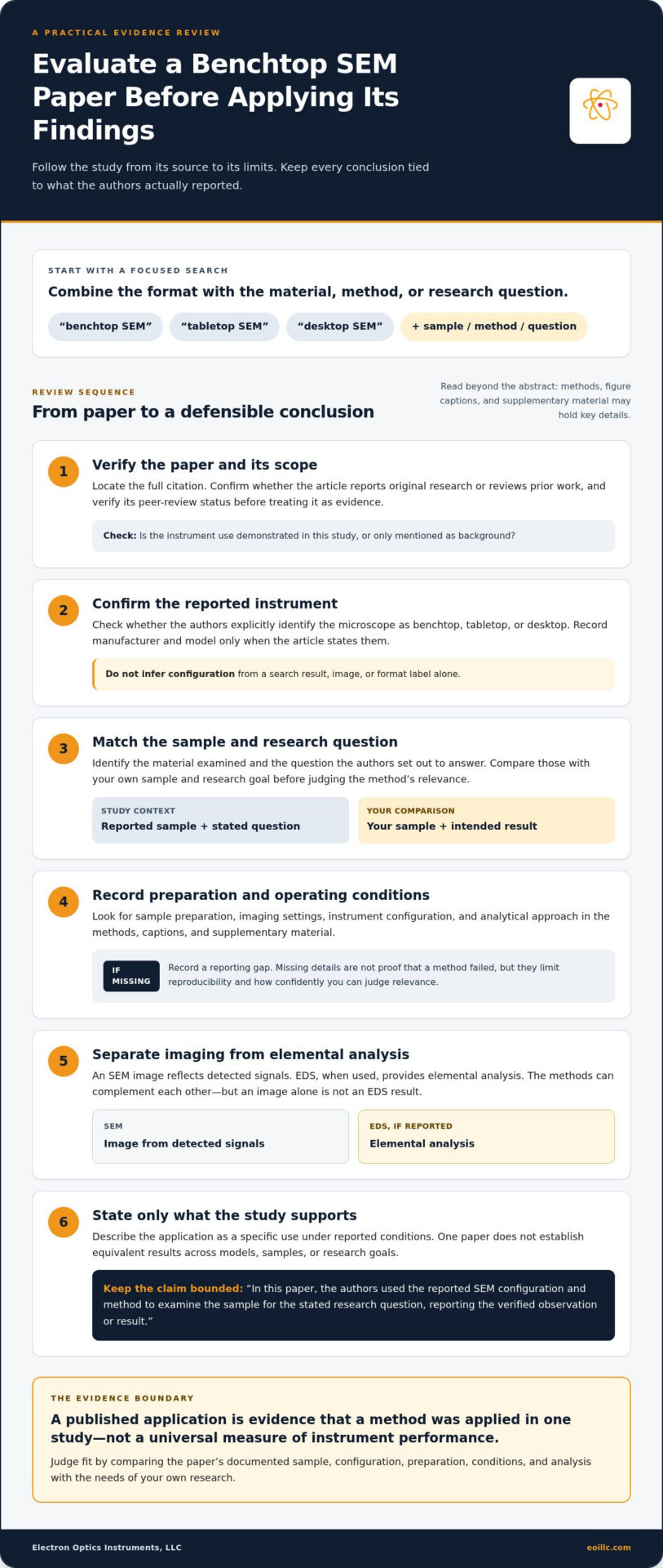
A consistent review process helps you decide whether a published method is relevant to your research. For each paper, use this checklist and record what the authors state rather than filling gaps with assumptions:
- Citation: Capture the complete title, authors, journal, publication date, and DOI or other identifier.
- Peer-review status: Verify the article type and publication record through the journal. Distinguish primary research from reviews or conference material.
- Instrument: Record the manufacturer and exact model, along with whether the authors identify it as benchtop, tabletop, or desktop.
- Sample: Note the material, condition, and preparation relevant to the study.
- Settings: Capture reported operating details, including accelerating voltage, detector, working distance, and vacuum mode.
- Method: Identify the imaging approach and any additional techniques, such as EDS.
- Results: Summarize only the observations and measurements the authors report.
- Limitations: Record caveats stated by the authors and note important methodological details that are absent.
Keep evidence types distinct. SEM images support claims about observed morphology or surface features under the reported conditions. EDS results support claims about elemental analysis, while other analytical techniques answer their own specific questions. If a paper reports both imaging and EDS, attribute each result to the method that produced it rather than treating an image as proof of composition.
Which instrument and operating details should readers capture?
Instrument model and operating conditions provide context for interpreting findings. Two papers are not directly comparable just because both use a benchtop SEM. Detector selection, voltage, working distance, vacuum mode, and sample protocols may differ. Record each detail when available. If a paper doesn’t state it, mark it as “not reported” rather than assuming a configuration or treating the omission as evidence of poor performance.
How sample preparation and limitations shape interpretation
Check the methods and supplementary material for coating, drying, sectioning, or other preparation steps. These can affect what the images reveal and how closely the protocol matches your samples. Look for discussion of charging, artifacts, resolution constraints, and study-specific caveats. Separate an author’s stated limitation from your own assessment, and cite the paper precisely when summarizing either.
For broader methodological context, consult the guide to advanced SEM techniques alongside this checklist. When reviewing peer-reviewed papers using benchtop SEM, a disciplined record of reported details and information gaps helps you compare studies without overstating what any one paper demonstrates.
How to Find and Compare Peer-Reviewed Benchtop SEM Papers
Broad searches for scanning electron microscopy can return studies using many instrument formats. Narrow the results by pairing a format term with the material, method, or research question you care about. Try “benchtop SEM” with a sample name, then repeat the search with “tabletop SEM” and “desktop SEM.” Add terms such as “EDS,” “surface imaging,” or a specific application to refine the results. Treat titles and snippets as discovery leads, not proof of a paper’s methods or findings.
Search terms and databases for focused literature discovery
Use scholarly indexes to identify citations, then follow links to the journal or publisher record and full text. Verify the article type and peer-review status through the journal record. Check the paper’s references for related studies and alternate terminology. Search an exact microscope manufacturer or model only when it appears in a paper or a reliable manufacturer record. For background before assessing studies, review a foundational overview of SEM principles.
Build a comparison matrix without overstating the evidence
A matrix helps make relevant similarities and differences visible without turning unrelated experiments into an instrument ranking. Use one row per paper and enter only details supported by its full text. Mark omissions as “not reported” to distinguish unknown information from a demonstrated limitation.
| Comparison field | What to record |
|---|---|
| Application and objective | The research question and the task the SEM was used to address |
| Sample and preparation | Material examined and stated preparation steps |
| Instrument identification | Benchtop, tabletop, or desktop designation, plus manufacturer and model if reported |
| Imaging and analytical methods | Imaging approach and any distinct methods, such as EDS |
| Results and limitations | Reported observations, author-stated caveats, and details not reported |
Compare papers first by research objective and sample, then by preparation, instrument details, and analytical output. Two studies may use the same format label yet address different questions under different conditions, so their findings shouldn’t be treated as a direct performance comparison. Use the matrix to identify evidence relevant to your application, not as a substitute for evaluating the full methods.
Use the comparison to turn literature findings into practical equipment questions for your lab. Discuss benchtop SEM options for your research requirements as you move from published methods to system evaluation.
From Published Evidence to a Benchtop SEM for Your Laboratory
Published methods become useful for equipment evaluation when you translate them into requirements for your laboratory. A paper may describe an approach relevant to your work, but its value depends on how closely its sample, imaging objective, and analytical method match your own. Separate directly relevant evidence from workflows that would need validation with your materials or research conditions.
Build a practical requirements list before comparing systems:
- Samples: Identify the materials, sample sizes, and preparation workflows your research involves.
- Imaging goals: Specify the features or structures you need to examine and the image outputs your work requires.
- Analytical needs: Decide whether imaging is sufficient or whether EDS or another analytical method is part of the research question.
- Throughput: Consider how frequently samples need to be examined and how the workflow fits laboratory operations.
- Users: Account for operator experience, training needs, and the number of people who will use the system.
- Ongoing support: Include service and maintenance requirements in your planning.
This framework keeps the decision anchored to the work rather than a format label. Compare benchtop and larger SEM formats against the same research requirements, considering what each setup needs to accomplish and how it fits the lab’s workflow. A broader discussion of how desktop SEMs have evolved can provide additional context, but published application examples should still be assessed against your samples and research questions.
Explore equipment and support for research workflows
Electron Optics Instruments offers the Cube II Benchtop SEM for evaluation against defined research requirements. Its mention here does not suggest that the system was used in any paper discussed. Start with your sample types, imaging objectives, and analytical needs, then consider how the equipment and workflow align.
Research workflows also depend on effective operation and ongoing instrument care. Electron Optics Instruments provides SEM service, maintenance, repairs, and on-site training. Include these considerations alongside imaging and EDS needs when planning for laboratory use.
Use peer-reviewed papers using benchtop SEM as evidence for specific methods, then convert relevant findings into questions about your research workflow. To discuss your laboratory requirements and explore benchtop SEM solutions, connect published evidence to the work your system needs to perform.
Turn Published Evidence Into a Practical Research Plan
Peer-reviewed papers using benchtop SEM can show how researchers apply these systems, but their value depends on the details: sample, instrument configuration, methods, and limitations. Treat each study as evidence for a defined application, then compare its conditions with your laboratory’s imaging goals, analytical needs, throughput, and users.
This approach also makes equipment evaluation more rigorous. Electron Optics Instruments brings more than 30 years of industry experience to SEM systems and support, including the Cube II Benchtop SEM, SEM service, maintenance, repairs, and on-site training. Assess the Cube II against your requirements, not as a claim about equipment used in the papers reviewed.
Explore benchtop SEM solutions for your research workflow and take the next step from published methods to a system aligned with your laboratory’s work. Start with a clear list of samples, imaging objectives, and analytical needs.
Frequently Asked Questions
Are there peer-reviewed papers using benchtop SEM?
Yes. Peer-reviewed papers report research using compact SEM formats, but verify each paper’s peer-review status and instrument type individually. Search records may use “benchtop,” “tabletop,” or “desktop,” and those terms alone don’t identify a specific microscope or configuration. Check the journal record and full paper before treating a result as evidence for an application or instrument capability.
What applications have researchers studied with benchtop SEM?
Applications vary by study, sample, and research question. Gunshot residue and cathodoluminescence are useful search leads, but a paper title alone doesn’t establish what the authors examined or found. Before describing either as a demonstrated application, verify the sample type, whether the article reports original research, the imaging and analytical methods, and the results in the full paper. Don’t extend a study’s findings beyond its reported scope.
How can I find peer-reviewed papers using a benchtop SEM?
Search scholarly indexes using “benchtop SEM,” “tabletop SEM,” or “desktop SEM” alongside a sample, method, or research question. For example, combine a format term with “EDS” or a material name. Review results on the journal or publisher page, confirm the article type and peer-review status, then inspect the full text and references. Search exact manufacturer or model names only when a reliable record supplies them.
Can benchtop SEM papers prove that an instrument will work for my samples?
No single paper can guarantee that an instrument will produce the same results with your samples. Published work demonstrates a method under particular conditions, including a defined sample, preparation protocol, instrument configuration, and research objective. Compare those conditions with your own and identify where further testing may be needed. A relevant paper can inform evaluation, but it isn’t universal proof of suitability or performance.
What details should I check in a benchtop SEM research paper?
Check the full citation, peer-review status, instrument designation, manufacturer and model, sample type, and preparation steps. Record operating details such as accelerating voltage, detector, working distance, and vacuum mode when reported. Separate image-based observations from EDS or other analytical results, and note the authors’ findings and limitations. Mark absent details as “not reported” rather than inferring settings or capabilities from the title or abstract.
Does benchtop SEM research always include EDS analysis?
No. SEM imaging and EDS are distinct methods, and a study may use SEM imaging without elemental analysis. SEM images are used to examine features captured by the selected imaging signal; EDS provides elemental information when that technique is performed and reported. Check the methods and results to see which analyses were conducted. Don’t interpret an SEM image alone as proof of sample composition.
How should I compare papers that use different SEM models?
Compare studies by research question, sample, preparation, operating conditions, imaging method, and analytical outputs, not by model names alone. Differences in detectors, settings, and workflows can affect what each study demonstrates. Build a comparison table and record unreported details explicitly. Use it to find papers relevant to your requirements, rather than ranking instruments based on experiments with different samples or methods.