
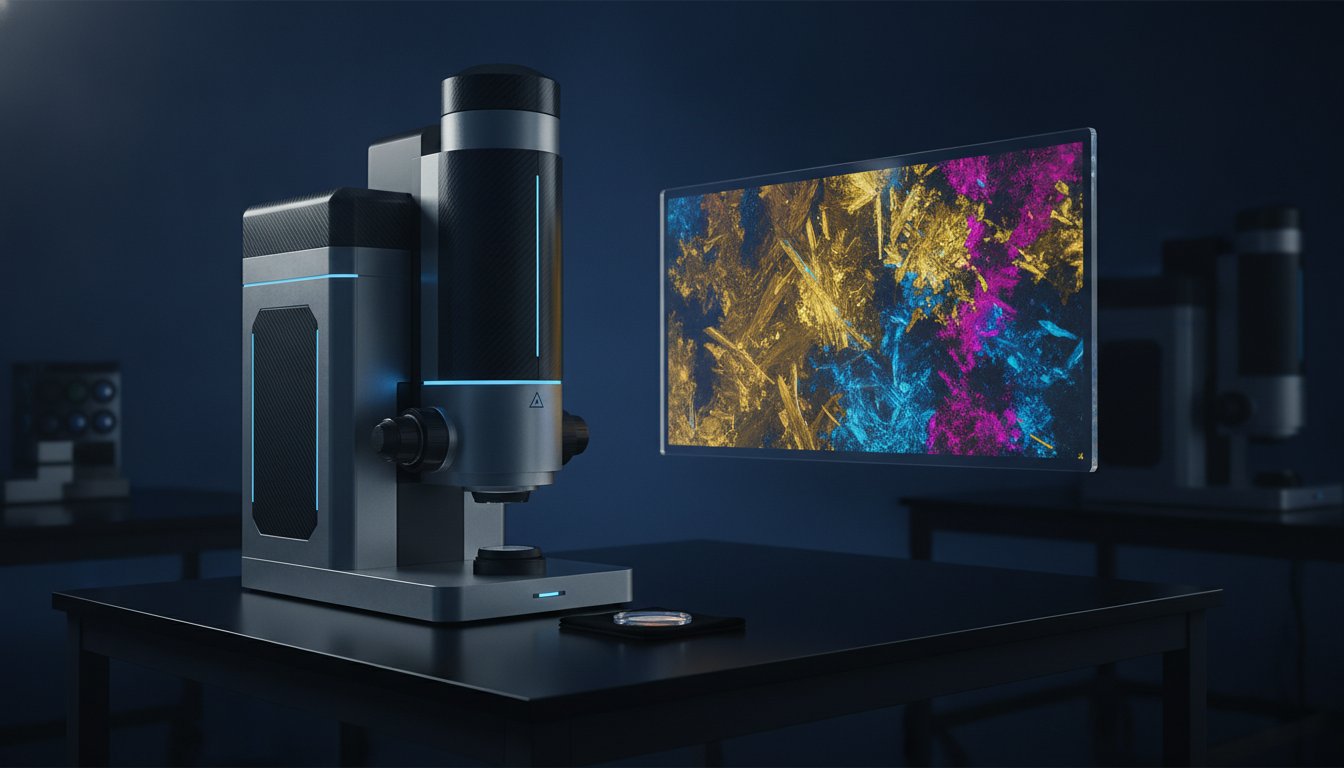
The era where high-fidelity mineralogical characterization required a dedicated, multi-room laboratory footprint has officially concluded. For years, industry professionals accepted the immense capital expenditure and infrastructure demands of floor-standing units as the unavoidable price of analytical precision. However, the contemporary landscape of geological sample analysis sem has undergone a fundamental transformation, driven by the integration of field-emission performance into compact, benchtop architectures. You likely recognize the logistical strain of sending samples to external facilities or managing the rigorous maintenance requirements of legacy systems.
It’s clear that the demand for rapid, on-site mineral identification is no longer a luxury but a strategic necessity in 2026. This guide demonstrates how modern benchtop technology, such as the Veritas HR and Cube II series, delivers the analytical precision once reserved for massive floor models. We’ll explore the technical mechanisms enabling high-resolution imaging of mineral textures and the deployment of advanced EDS systems for accurate elemental mapping. By the end of this professional briefing, you’ll understand how to achieve superior petrographic data while significantly reducing your cost per sample analysis.
Key Takeaways
- Understand the paradigm shift from traditional optical petrography to advanced sub-micron morphological and chemical characterization using modern electron optics.
- Identify the specific roles of Backscattered Electron (BSE) and Secondary Electron (SE) imaging in enhancing the accuracy of geological sample analysis sem.
- Acquire specialized protocols for preparing petrographic thin sections and polished blocks, including methods for mitigating charging effects on non-conductive mineral samples.
- Compare the operational throughput and reduced infrastructure requirements of benchtop SEM systems against traditional floor-standing analytical models.
- Learn how to align specific SEM configurations with specialized geological disciplines to optimize data acquisition and ensure sustained laboratory uptime.
The Evolution of SEM in Geological Analysis
A Scanning Electron Microscope (SEM) serves as a sophisticated, non-destructive analytical platform designed for sub-micron morphological and chemical characterization of mineral specimens. In the specialized field of geological sample analysis sem, this technology acts as the primary bridge between macroscopic observation and nanometre-scale mineralogy. By 2026, the industry has transitioned away from the rigid constraints of centralized, floor-standing units. We’re seeing a decisive shift toward decentralized infrastructure that prioritizes immediate data acquisition without sacrificing the rigorous vacuum stability or electron optic precision required for complex geological specimens.
Traditional methods often fall short when dealing with the intricate textures of modern exploration targets. SEM has surpassed optical petrography because it provides a level of detail that light-based systems simply can’t reach. Recent advancements in 2026 have introduced enhanced field-emission stability and automated vacuum management systems. These innovations ensure that even the most volatile or porous geological samples remain stable during high-resolution imaging sequences. This evolution reflects a broader commitment to technical integration and logistical excellence within the geosciences.
Limitations of Traditional Optical Microscopy
Optical microscopy remains a foundational skill, yet its technical boundaries are increasingly problematic for high-stakes research. The inherent depth of field constraints make it nearly impossible to accurately assess 3D grain morphology or complex fracture networks in thick sections. At the nanometer scale, resolution barriers prevent the detailed micro-porosity analysis essential for unconventional reservoir characterization. Perhaps most critically, optical systems don’t allow for real-time elemental mapping. Researchers must move samples to secondary instruments, which increases the risk of contamination and delays critical decision-making.
The Rise of Benchtop SEM in Geoscience
The emergence of high-performance hardware, such as the Cube II Benchtop SEM, has effectively democratized advanced analytical capabilities. These systems match the performance of traditional floor models while occupying a fraction of the space. By integrating high-brightness filaments, these instruments provide superior imaging of non-conductive rock samples that previously required extensive preparation. Portability is the final piece of the puzzle. The ability to deploy these systems in remote mining facilities or mobile exploration labs ensures that geological sample analysis sem can occur at the point of extraction, streamlining the path from discovery to production.
Microstructural and Elemental Characterization Techniques
The efficacy of geological sample analysis sem relies on the precise calibration of electron-matter interactions to resolve complex mineralogical puzzles. We achieve this through the strategic application of diverse signal detection modes, each revealing distinct physical and chemical properties of the specimen. While secondary electron (SE) imaging provides the topographic clarity necessary to assess surface morphology and the progression of chemical weathering, it’s the integration of backscattered electron detection and energy dispersive spectroscopy that forms the backbone of modern petrographic characterization. This multi-modal approach ensures that no microstructural detail is overlooked during the investigative process.
BSE Imaging and Atomic Number Contrast
Backscattered electron (BSE) imaging utilizes the elastic scattering of electrons to generate images based on atomic number (Z) contrast. This technique is indispensable for distinguishing between heavy and light mineral phases in complex ore samples, where bright intensities correlate directly with higher average atomic numbers. It allows for the immediate identification of micro-inclusions and subtle compositional zoning patterns within igneous crystals that optical systems inevitably miss. Furthermore, researchers can optimize contrast parameters to facilitate rapid differentiation between carbonate and silicate matrices, ensuring that high-density accessory minerals, such as zircon or monazite, are clearly visible against a lower-density quartz-feldspar groundmass.
High-Resolution EDS Mapping in 2026
The analytical standard for 2026 demands the rapid acquisition of multi-element maps to support large-scale sedimentary provenance studies and economic resource estimation. Modern EDS (Energy Dispersive Spectroscopy) Systems integrated into the Genesis and Veritas series have significantly reduced detector dead-time, allowing for the detection of trace elements in sulfide minerals with unprecedented speed. These advancements enable the generation of detailed chemical heat maps that reveal the spatial distribution of critical metals, which is essential for understanding ore genesis and mineral liberation.
By transitioning from raw spectral data to automated mineralogy workflows, laboratories can now produce comprehensive modal mineralogy reports that quantify mineral abundance and grain size distributions. This systematic approach ensures that petrographic data is both reproducible and statistically significant. If your facility requires enhanced characterization throughput, the technical capabilities of the Veritas Series SEM provide a clear path toward industrial-grade analytical excellence. The synergy between high-sensitivity detectors and intuitive software allows for the seamless transformation of complex electron signals into actionable geological intelligence, bridging the gap between raw data and strategic insight.
Overcoming Challenges in Geological Sample Preparation
The integrity of geological sample analysis sem is fundamentally determined by the quality of initial specimen preparation. Even the most advanced electron optics cannot compensate for poor surface geometry, residual surface deformation, or volatile contamination. While previous sections discussed the analytical capabilities of modern systems, those results are only as reliable as the preparation protocols used to stabilize the specimen. Professional laboratories must implement rigorous standards to ensure that the mineralogical data extracted is representative of the original geological context without artifacts introduced during the cutting or mounting phases.
Success in this area requires a meticulous approach to material science, specifically regarding how geological materials react under high-vacuum conditions. Managing sample outgassing is critical for porous sedimentary rocks and moisture-rich specimens. High-vacuum environments can cause sample degradation or vacuum instability if volatiles aren’t properly removed through pre-analysis desiccation or controlled heating. For microfossils and particulate matter, specialized mounting techniques using conductive carbon adhesives ensure stability under the electron beam without obscuring delicate morphological features essential for taxonomic identification.
Cutting, Mounting, and Polishing Protocols
Achieving absolute planarity is non-negotiable for accurate EDS quantitation and electron backscatter diffraction (EBSD) analysis, where tilt angles and surface smoothness directly influence signal yield. Technicians must select mounting resins that exhibit high vacuum stability and superior edge retention to prevent separation between the mineral grain and the matrix during the polishing process. The final stages of preparation should utilize colloidal silica to produce a deformation-free surface. This ensures that the crystalline lattice remains intact, which is a prerequisite for high-resolution characterization in advanced series like the Veritas HR.
Coating and Conductivity Management
Non-conductive geological materials, such as silicates and carbonates, require precise conductivity management to prevent electron accumulation and subsequent image distortion. Carbon coating remains the industry standard for EDS and WDS applications because it offers minimal interference with characteristic X-ray signals. Conversely, gold or palladium coatings are preferred for high-resolution secondary electron imaging where surface topography is the primary focus. A sputter coater is an essential laboratory instrument that deposits a nanometer-scale conductive layer onto the sample surface to facilitate stable electron interaction and prevent thermal damage during geological sample analysis sem. For sensitive or rare samples that cannot be coated, utilizing low-vacuum or variable pressure modes provides a viable alternative by using gas molecules to neutralize surface charge.
The Strategic Advantage of Benchtop SEM in Geosciences
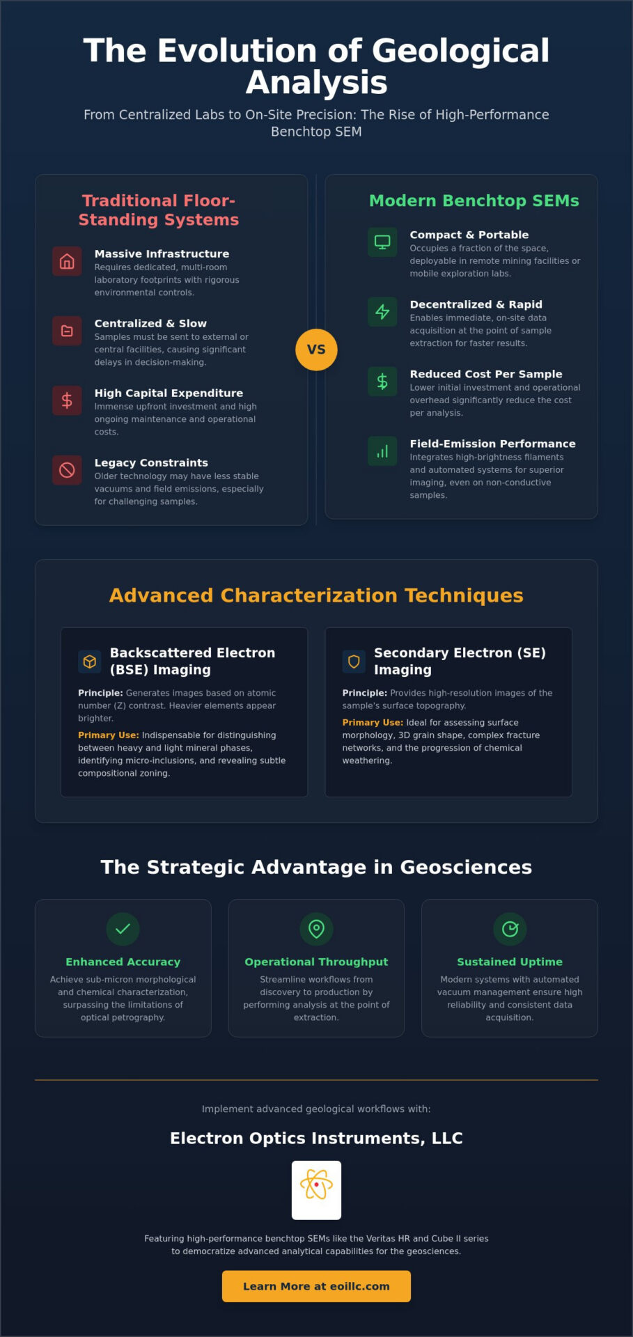
The transition toward benchtop systems represents a paradigm shift in how research institutions and industrial partners manage their analytical assets. Traditional floor-standing units, while powerful, often create logistical bottlenecks due to their extreme sensitivity to environmental noise and high maintenance requirements. In contrast, the current generation of benchtop systems for geological sample analysis sem provides equivalent resolution without the need for specialized vibration isolation or dedicated cooling loops. This operational agility allows organizations to deploy high-end characterization tools directly within active exploration facilities or multi-user university labs where floor space is a premium. By decentralizing analytical power, teams can achieve higher throughput and maintain tighter control over their data pipelines.
Infrastructure requirements for benchtop units are remarkably lean compared to their larger counterparts. Most systems operate on standard electrical circuits and require minimal environmental controls, avoiding the climate-stabilized enclosures demanded by legacy hardware. When evaluating the Total Cost of Ownership in 2026, it’s essential to consider the reduced frequency of Preventative Maintenance Visits and the lower cost of consumables like filaments. This scalability makes it feasible for a single department to operate multiple units, effectively increasing overall capacity and ensuring that critical research isn’t delayed by a single point of failure in a centralized facility.
ROI and Economic Justification
Analyzing the electron microscope cost reveals that the initial investment in a benchtop system often pays for itself within eighteen to twenty-four months when compared to the escalating fees and shipping costs associated with third-party analytical laboratories. In-house analysis eliminates the logistical delays inherent in external outsourcing and allows for iterative testing. This rapid turnaround is a decisive factor in exploration projects where immediate mineralogical data can influence drilling strategies and real-time resource estimation. The long-term impact on project budgets is substantial, as reliability and lab uptime are maximized through simplified internal components and robust engineering.
Ease of Use and Training Requirements
Modern software interfaces have revolutionized the user experience, significantly lowering the barrier to entry for non-specialists. Automated alignment and focus routines ensure that high-quality data is captured consistently, regardless of the operator’s prior experience level. This shift reduces the reliance on a single, dedicated SEM operator and allows geologists to interact directly with their samples in a more intuitive environment. On-site training programs act as a catalyst for immediate research productivity, enabling teams to transition from installation to active characterization within days. For organizations aiming to enhance their technical integration, investing in the Veritas Series SEM through Electron Optics Instruments, LLC provides a clear path toward localized analytical authority and superior standards in mineralogical research. If your laboratory requires a transition to high-precision benchtop technology, explore our advanced hardware configurations to optimize your research throughput.
Implementing Advanced Geological Workflows with Electron Optics Instruments, LLC
The successful integration of high-throughput workflows into a geoscience facility requires a meticulous alignment between research objectives and hardware specifications. We recognize that the requirements of a paleontology lab, which may prioritize delicate surface morphology, differ fundamentally from the quantitative rigor demanded by economic geology. Selecting the appropriate platform for geological sample analysis sem isn’t merely a procurement decision; it’s a strategic move that defines the analytical boundaries of your institution for the next decade. By coordinating specialized hardware with specific mineralogical goals, Electron Optics Instruments, LLC ensures that your laboratory data acquisition remains both relevant and statistically robust.
Future-proofing a laboratory in 2026 involves more than just selecting high-resolution optics. It requires a visionary approach to technical integration where the instrument, the operator, and the support infrastructure function in total synergy. Whether your focus is on unconventional reservoir characterization or large-scale mining operations, the ability to generate reproducible results under varying environmental conditions is paramount. This stability is the hallmark of a seasoned global innovator, providing the reliability needed to push the boundaries of what’s possible in advanced hardware systems.
Customized Solutions for Geological Labs
The Cube II Benchtop SEM provides a robust solution for field-based mineral identification, offering the portability required for remote exploration without compromising on the vacuum stability discussed in earlier sections. For research environments focused on nano-scale characterization and high-resolution imaging, the Veritas series, including the Veritas HR and Veritas FE models, offers superior beam brightness and stability. When capital budgets are constrained, integrating Refurbished SEM Units allows departments to expand their analytical capacity while maintaining the high standards of accuracy associated with new hardware. This tiered approach ensures that every laboratory can access professional-grade electron optics.
Maintenance and Long-Term Reliability
Preserving the precision of these instruments over a multi-year lifecycle depends on rigorous SEM maintenance. Consistent beam stability and detector sensitivity are only achievable through scheduled Preventative Maintenance Visits, which mitigate the risk of unexpected research downtime. Electron Optics Instruments, LLC brings a 30-year legacy of technical excellence to this supporting role, acting as a reliable partner for national geoscience labs and private industrial firms alike. Service contracts provide a visionary safeguard, ensuring your facility remains at the cutting edge of geological sample analysis sem while your internal processes stay focused on discovery and production. By choosing a partner that values international capability and meticulousness, you ground your laboratory in the practical realities of logistical excellence.
Advancing Mineralogical Discovery into 2026
The paradigm shift toward decentralized, high-performance electron optics has fundamentally altered the landscape of mineralogical research. We’ve detailed how benchtop architectures now provide the analytical depth required for complex characterization without the infrastructure burdens or capital intensity of legacy systems. By integrating the preparation protocols and EDS mapping techniques explored in this guide, your facility can achieve a level of precision previously reserved for the most advanced centralized hubs. This transformation ensures that geological sample analysis sem remains a resilient cornerstone of both industrial exploration and academic research as we move into a new era of technical integration.
As the sole US distributor for EmCraft SEMs with more than 30 years of specialized expertise, Electron Optics Instruments, LLC provides the technical integration, comprehensive service, and on-site training necessary to sustain long-term laboratory uptime. Our commitment to meticulousness and international capability ensures your team is equipped for the rigorous demands of modern petrography and sub-micron characterization. Explore the Cube II and Veritas SEM series for your geological lab to establish a new standard of accuracy in your characterization workflows. We’re ready to help you push the boundaries of what’s possible in your analytical environment.
Frequently Asked Questions
Why is SEM preferred over optical microscopy for geological sample analysis?
Scanning electron microscopy provides superior depth of field and sub-micron resolution that far exceeds the physical limitations of light-based systems. While optical tools are restricted by the wavelength of light, an SEM utilizes electron beams to resolve nanometre-scale features such as micro-porosity and complex grain boundaries. This capability is essential for modern geological sample analysis sem, where identifying subtle mineralogical textures is critical for accurate reservoir characterization and ore grade estimation.
Can benchtop SEMs handle the high-vacuum requirements for geological specimens?
Modern benchtop units, including the Cube II and Veritas series, utilize sophisticated turbomolecular pump systems that achieve the rigorous high-vacuum levels necessary for stable geological imaging. These systems effectively manage the outgassing common in porous sedimentary rocks and moisture-sensitive minerals. This technical integration ensures that decentralized laboratories maintain the same analytical integrity as traditional centralized facilities while operating within a significantly smaller physical footprint.
What is the primary difference between BSE and SE imaging in geology?
Backscattered Electron (BSE) imaging identifies compositional variations based on atomic number contrast, whereas Secondary Electron (SE) imaging focuses on surface topography and morphology. In a geological context, BSE allows researchers to distinguish between mineral phases like pyrite and quartz based on their brightness. SE imaging is primarily utilized to examine weathering patterns, fracture networks, and the three-dimensional structure of microfossils or crystalline surfaces.
How do I prevent my geological samples from charging during SEM analysis?
Charging is mitigated by applying a conductive nanometre-scale coating of carbon or gold or by utilizing variable pressure (low-vacuum) modes. Carbon coating is the professional standard for EDS applications because it doesn’t interfere with characteristic X-ray signals. If a sample is too fragile for coating, variable pressure systems neutralize surface charge by introducing a small amount of gas into the chamber, which allows for stable imaging of non-conductive silicates.
Is EDS analysis on a benchtop SEM accurate enough for mineral identification?
Current energy dispersive spectroscopy systems integrated into benchtop platforms deliver professional-grade quantitative data suitable for definitive mineral identification. These detectors provide the spectral resolution required to distinguish between complex solid-solution series and detect trace elements in sulfide minerals. When paired with automated mineralogy software, these instruments generate statistically significant modal mineralogy reports that meet the rigorous standards of both industrial exploration and academic research.
What are the infrastructure requirements for installing a benchtop SEM in a geology lab?
Infrastructure requirements are minimal, typically involving a standard electrical outlet and a stable, vibration-resistant laboratory bench. Unlike floor-standing units, these systems don’t require dedicated cooling water loops, specialized nitrogen lines, or reinforced flooring. This simplicity allows for rapid deployment in mobile exploration units or remote mining facilities, providing a strategic advantage for projects requiring immediate on-site geological sample analysis sem results.
How often does an SEM used for geological samples require preventative maintenance?
Geological laboratories should schedule professional preventative maintenance visits at least once per year to ensure sustained beam stability and detector sensitivity. High-throughput facilities or those analyzing particularly dusty particulate samples may benefit from semi-annual servicing to prevent aperture contamination. Regular maintenance preserves the longevity of high-brightness filaments and ensures the vacuum system remains at peak performance, which is essential for maintaining rigorous analytical standards.
Can I use an SEM to analyze micro-porosity in reservoir rocks?
High-resolution models like the Veritas HR or Veritas FE are specifically engineered to resolve the complex micro-porosity networks found in unconventional reservoir rocks. These instruments allow geologists to quantify pore throat distributions and connectivity at the nanoscale, which is vital for predicting fluid flow and storage capacity. By utilizing advanced electron optics, researchers can visualize the intricate relationship between organic matter and mineral matrices in shale and tight gas formations.