

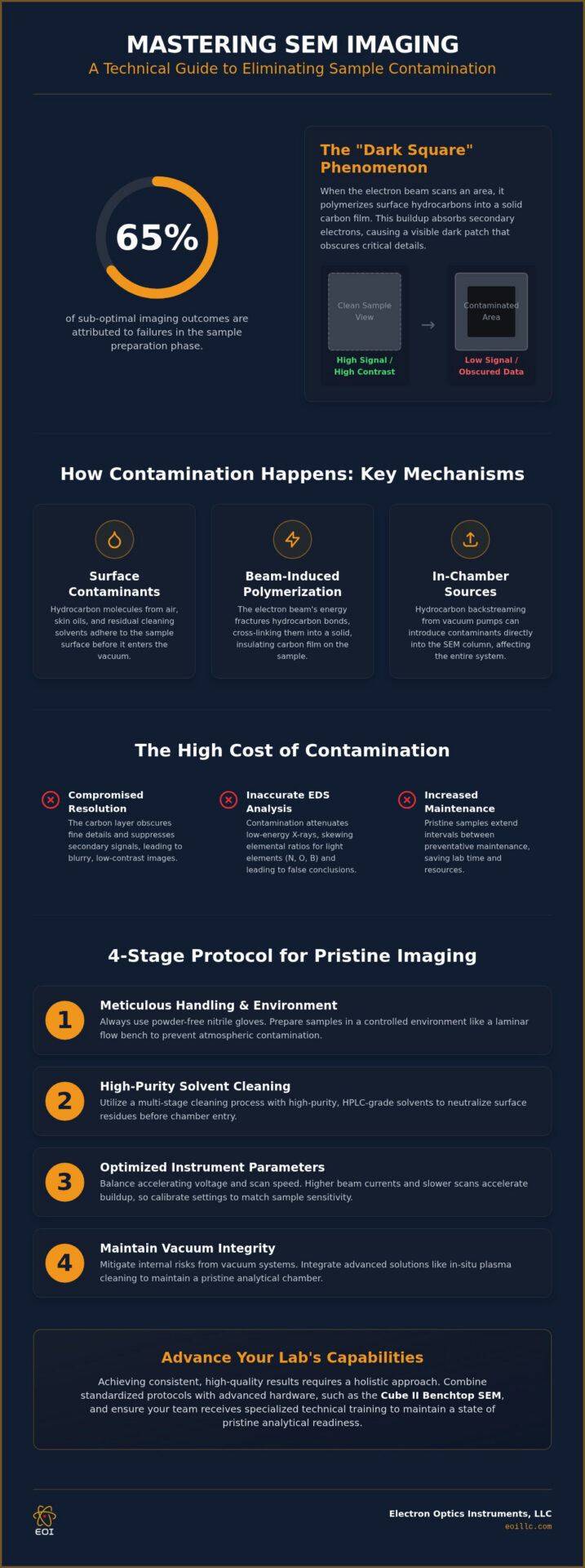
Did you know that an estimated 65% of sub-optimal imaging outcomes in industrial laboratories are directly attributed to failures in the sample preparation phase? For many researchers, the frustration of observing dark carbonaceous patches obscure a critical microstructure is a recurring obstacle that compromises both resolution and analytical accuracy. It’s clear that achieving the highest standards of imaging excellence requires more than just high-end hardware; it demands a rigorous commitment to avoiding sample contamination in sem through methodical precision and technical discipline.
This guide provides the technical expertise necessary to master the professional protocols and instrument settings required to eliminate carbonaceous buildup while preserving your vacuum integrity. By implementing these standardized preparation techniques, your team can secure crisp, high-contrast images at extreme magnifications and significantly extend the intervals between preventative maintenance visits. We’ll examine the critical intersection of in-situ plasma cleaning, optimized accelerating voltages, and the advanced capabilities of systems like the Cube II Benchtop SEM to ensure your laboratory maintains a state of pristine analytical readiness.
Key Takeaways
- Identify the fundamental mechanisms of carbonaceous buildup, specifically the polymerization of surface hydrocarbons that results in the “dark square” phenomenon during high-magnification scans.
- Establish a multi-stage sample preparation workflow utilizing high-purity HPLC-grade solvents to ensure surface residues are neutralized prior to vacuum chamber entry.
- Recognize and mitigate internal contamination risks, including hydrocarbon backstreaming from vacuum pumping systems, to preserve the long-term integrity of the SEM column.
- Optimize instrument parameters by balancing accelerating voltages and resolution requirements to minimize beam-induced buildup on sensitive industrial materials.
- Integrate advanced hardware solutions like the Cube II Benchtop SEM with specialized technical training to standardize protocols for avoiding sample contamination in sem across your entire laboratory.
Understanding the Mechanisms of SEM Sample Contamination
The integrity of imaging within a Scanning Electron Microscope depends heavily on the purity of both the vacuum environment and the specimen surface. Carbonaceous contamination represents a critical challenge for researchers; it’s the chemical result of hydrocarbon molecules undergoing polymerization under the direct influence of the primary electron beam. These organic contaminants aren’t always inherent to the sample’s composition. They frequently originate from atmospheric exposure, the transfer of skin oils during manual handling, or residual solvents left behind by sub-optimal cleaning protocols. When the electron beam interacts with these mobile organic molecules, it facilitates a phase change, fixing them to the surface as a resilient, solid carbon film.
The sample substrate itself often acts as a reservoir for these molecules, as porous or complex surfaces can harbor trapped organics that migrate toward the beam’s impact point through surface diffusion. This accumulation manifests visually as the “Dark Square” phenomenon. As the electron probe scans a specific area, the localized concentration of carbon increases in thickness. This newly formed layer effectively absorbs secondary electrons that would otherwise escape the surface to reach the detector, leading to a quantifiable reduction in signal-to-noise ratios. This dark patch is far more than a mere visual artifact; it’s a physical barrier that obscures fine topographic details and suppresses the emission of secondary signals. Establishing a rigorous framework for avoiding sample contamination in sem is essential for any facility that prioritizes the acquisition of reproducible, high-contrast data at the nanometer scale.
The Role of the Electron Beam in Polymerization
The speed of carbon deposition is fundamentally linked to the beam’s current density and the total residence time of the probe on the specimen. Primary electrons and the resulting secondary electrons provide the necessary energy to fracture molecular bonds in surface-adsorbed hydrocarbons, which then facilitates the rapid cross-linking of carbon chains. Electron-beam-induced deposition (EBID) acts as the primary driver behind this unwanted material accumulation during the imaging process. Higher beam currents and slower scan speeds typically accelerate this transformation, making methodical instrument calibration a prerequisite for maintaining a clean and accurate imaging field.
Impact on Analytical Accuracy and EDS
Surface contamination poses a significant threat to the validity of quantitative EDS analysis. As the carbonaceous layer thickens, it begins to attenuate low-energy X-rays emitted from the underlying material, specifically affecting the detection of light elements such as nitrogen, oxygen, and boron. This absorption phenomenon skews elemental ratios, which can lead to highly inaccurate stoichiometric conclusions in industrial failure analysis or materials characterization. Maintaining pristine surfaces is a non-negotiable requirement for accurate elemental mapping, as even a nanometric layer of carbon can significantly alter the detected spectra. Achieving professional results requires a holistic approach to avoiding sample contamination in sem, ensuring the vacuum and the specimen remain free of organic interference.
Effective Sample Preparation and Cleaning Protocols
The transition from theoretical understanding to the practical execution of multi-stage cleaning protocols is essential for neutralizing the mechanisms of SEM contamination. While instrument outgassing and pumping systems are recognized contributors, the specimen itself often acts as the primary vehicle for volatile organic compounds entering the vacuum chamber. Meticulous handling protocols, including the mandatory use of powder-free nitrile gloves, are required to prevent the transfer of sebaceous oils that catalyze carbonaceous buildup. Establishing a controlled preparation environment, ideally within a clean-room or a dedicated laminar flow bench, remains a non-negotiable standard for any facility dedicated to avoiding sample contamination in sem.
Storage solutions must mirror this meticulousness to ensure long-term imaging success. Prepared specimens should be housed in desiccators or specialized vacuum pellicles to mitigate the risk of re-contamination from ambient atmospheric particulates. These proactive measures ensure that the surface remains pristine between the preparation and imaging phases, safeguarding the integrity of your analytical data and preventing the introduction of new hydrocarbons into the sensitive vacuum environment.
Solvent Cleaning and Ultrasonic Bathing
Chemical cleaning strategies must prioritize the use of high-purity, HPLC-grade solvents to eliminate the risk of non-volatile residues that lower-grade chemicals often leave behind. A standardized workflow typically involves a primary degreasing phase with acetone, followed by a secondary rinse in high-purity ethanol or isopropanol to displace dissolved organic films. For robust substrates such as industrial metals or ceramics, a 10-minute ultrasonic bath provides the necessary kinetic energy to dislodge surface-adsorbed particulates. It’s critical that drying is performed using filtered, oil-free compressed nitrogen; allowing solvents to air-dry often precipitates dissolved contaminants back onto the specimen surface, creating a localized film that obscures fine detail.
Advanced Surface Treatment: Plasma Cleaning
Advanced surface treatment through oxygen or argon plasma offers an atomic-level solution by chemically volatilizing residual hydrocarbon chains that chemical solvents cannot reach. Integrating these technologies into your SEM operation effectively neutralizes the primary drivers of electron-beam-induced deposition. In-situ plasma cleaners provide the advantage of treating the sample and the chamber environment simultaneously, whereas ex-situ units facilitate high-throughput preparation for large batches before loading. To maintain these rigorous standards of vacuum purity and ensure your instrument performs at its peak, consider the value of professional preventative maintenance to address long-term hydrocarbon accumulation within the column.
Mitigating In-Chamber and Vacuum System Contamination
Meticulous sample preparation represents only one half of the analytical equation; the internal architecture of the microscope often harbors latent sources of organic interference that can undermine even the most rigorous cleaning protocols. While conventional wisdom suggests that vacuum systems are sterile, closed loops, the reality is that instrument outgassing and mechanical wear can introduce a steady stream of hydrocarbons into the imaging environment. Achieving a truly pristine state for avoiding sample contamination in sem requires a comprehensive understanding of how internal components, from the electron column to the specimen stage, interact with the vacuum. Overlooking these internal factors often leads to chronic imaging artifacts that persist regardless of how thoroughly a specimen is degreased.
Stage hygiene is a critical, yet frequently neglected, component of laboratory management. Sample holders, stubs, and the stage itself act as cumulative reservoirs for oils and particulates if they aren’t subjected to the same decontamination standards as the samples they carry. The presence of high-vapor-pressure materials, such as certain adhesives or inadequately degassed polymers, can initiate a cascade of outgassing events that compromise vacuum stability and increase the rate of carbonaceous buildup during long-duration scans.
Vacuum System Maintenance and Oil Management
Modern desktop SEM platforms have largely transitioned to dry, oil-free scroll pumps to eliminate the risk of hydrocarbon backstreaming, a phenomenon where pump oil vapors migrate against the pressure gradient and into the chamber. In systems still utilizing rotary vane pumps, the failure of anti-suckback valves or the degradation of pump oil can lead to significant contamination. Identifying these issues early is vital; a sudden increase in the “dark square” effect or a noticeable shift in vacuum recovery times often indicates that the system is contributing to specimen impurity. Consistent SEM preventative maintenance is the most effective strategy for ensuring that seals remain airtight and that the pumping infrastructure doesn’t become a primary source of carbon buildup.
Chamber Hygiene and Component Cleaning
Maintaining chamber hygiene involves a methodical approach to component care, particularly regarding O-rings and seals. While these components are essential for vacuum integrity, the application of excessive vacuum grease creates a significant source of volatile organics that the electron beam can easily polymerize. SOPs should dictate that O-rings are cleaned with lint-free wipes and only lightly lubricated with high-grade, low-vapor-pressure fluorinated greases. Furthermore, any tools used within the chamber, such as tweezers or mounting jigs, must be dedicated exclusively to SEM work and stored in a contaminant-free environment to prevent the accidental introduction of skin oils or industrial lubricants into the sensitive vacuum workspace.
Optimizing SEM Settings to Reduce Buildup During Imaging
Achieving imaging excellence requires a strategic calibration of instrument parameters to mitigate the kinetic energy that drives carbonaceous deposition. While high accelerating voltages are often prioritized for achieving maximum resolution, they simultaneously increase the rate of hydrocarbon polymerization on the specimen surface. Establishing an operational equilibrium between these competing requirements is a cornerstone of professional protocols for avoiding sample contamination in sem. By utilizing lower accelerating voltages, typically below 5 kV, operators can effectively reduce the interaction volume of the electron beam with surface-adsorbed organics, thereby preserving the structural integrity of beam-sensitive materials.
Strategic scanning techniques, such as the implementation of “Area of Interest” modes, allow for focused analysis while minimizing the total electron dose delivered to the surrounding topography. This targeted approach prevents the premature degradation of the wider field of view, ensuring that high-magnification captures remain free of the dark-square artifacts discussed in previous sections. For long-duration analytical tasks, the integration of active de-contamination hardware becomes essential to maintain a stable imaging environment that doesn’t compromise data reproducibility.
Voltage and Current Optimization
Refining the beam’s spot size and current is critical when transitioning from general topographical surveys to high-precision analytical tasks. Larger spot sizes, while beneficial for signal intensity, deliver a higher density of electrons that can rapidly fix contaminants to the surface. It’s vital to remember that an inverse relationship exists between scan speed and localized contamination because slower scan rates provide more time for mobile hydrocarbons to diffuse into the beam’s path and undergo polymerization. Operators should prioritize faster scan speeds for initial navigation, reserving slow, high-definition scans only for the final image acquisition phase to keep surfaces pristine.
Cryo-Trapping and Cold Finger Utility
In high-vacuum environments, the implementation of liquid nitrogen cold traps, or “cold fingers,” provides a physical mechanism for sequestering residual hydrocarbons. These surfaces are maintained at cryogenic temperatures, effectively pulling volatile organic molecules out of the vacuum and onto the trap before they can interact with the primary electron beam. This technique is particularly valuable during extended EDS mapping or EBSD analysis, where the specimen is subjected to prolonged beam exposure. When your research demands the highest levels of vacuum purity, it’s time to invest in active de-contamination hardware for your Veritas Series SEM to ensure consistent, artifact-free results. To explore how advanced hardware can streamline your laboratory’s workflow, view our full range of Veritas systems and support services.
Professional Solutions: Advanced Instruments and Training
While mastery of cleaning protocols and instrument settings is foundational, the long-term success of an analytical laboratory often hinges on the integration of advanced hardware and institutionalized knowledge. Modern systems are increasingly engineered to mitigate the human and environmental variables that contribute to specimen impurity. Transitioning to professional-grade solutions ensures that the technical discipline practiced by individual operators is supported by the inherent reliability of the instrument itself. This synergy between human expertise and hardware precision is the final requirement for maintaining a world-class imaging facility.
Customized solutions for specialized industrial failure analysis require more than just standard equipment; they demand a holistic approach to vacuum integrity. By utilizing instruments that prioritize clean operation from the design phase, laboratories can focus on data acquisition rather than constant troubleshooting. Establishing these high standards is the most effective way of avoiding sample contamination in sem, ensuring that every scan delivers the maximum possible resolution without the interference of organic artifacts.
Cube II and Genesis: Engineered for Precision
The Cube II Benchtop SEM represents a significant advancement in integrated vacuum management, utilizing automated sequences to ensure that chamber venting and loading cycles minimize atmospheric exposure. This level of automation reduces the window of opportunity for airborne hydrocarbons to enter the system, a critical factor in avoiding sample contamination in sem during high-throughput industrial workflows. For routine analysis, the Genesis Tabletop SEM provides a robust and user-friendly platform that maintains high-performance standards in a compact footprint. For facilities focused on high-resolution nanotechnology applications, the Veritas Series offers the expanded chamber capacity and specialized optics necessary for characterizing large-scale industrial components at the nanometer scale.
Maximizing Longevity Through Service and Education
Establishing lab-wide Standard Operating Procedures (SOPs) is simplified through specialized SEM training. These on-site programs act as a strategic investment in data integrity, ensuring that all staff members are proficient in the meticulous handling and cleaning standards required to preserve vacuum health. Beyond education, annual service contracts and Preventative Maintenance Visits provide a critical defense against the chronic, cumulative contamination that can occur within an electron column over time. Professional vacuum repair and maintenance ensure that internal seals, apertures, and pumping systems are operating at peak efficiency. For laboratories seeking to restore their instruments to factory-standard purity, contacting Electron Optics Instruments for specialized technical support is the most reliable path to ensuring equipment longevity and analytical accuracy.
Securing Analytical Excellence Through Technical Discipline
Transitioning from reactive cleaning to a proactive, holistic strategy is the only way to ensure reproducible imaging at the nanometer scale. By integrating multi-stage solvent purification with advanced instrument calibration, your laboratory can eliminate the carbonaceous artifacts that compromise data integrity. This methodical approach to avoiding sample contamination in sem transforms the microscope from a sensitive tool into a reliable cornerstone of industrial innovation. Meticulousness in sample handling and vacuum maintenance doesn’t just improve image contrast; it preserves the functional lifespan of your most critical hardware.
Electron Optics Instruments brings over 30 years of electron microscopy expertise to your facility as the sole US distributor for the EmCraft Cube II and Genesis series. We provide comprehensive service contracts for all major SEM brands, ensuring your vacuum systems remain pristine through expert-led preventative maintenance and specialized training. Explore our range of high-performance Benchtop SEMs and Service Solutions to elevate your laboratory’s capabilities and standardize your protocols. Your commitment to technical precision today secures the breakthrough discoveries of tomorrow.
Frequently Asked Questions
What are the most common signs of sample contamination in an SEM image?
The primary indicators include a visible darkening of the scanned region, often referred to as the “dark square” phenomenon, and a gradual loss of fine surface detail. You might also notice a decrease in the secondary electron signal, which leads to lower contrast and blurred edges at high magnifications. These artifacts occur when the electron beam polymerizes organic molecules on the surface, creating a carbon film that obscures the underlying topography.
Can I use regular rubbing alcohol to clean my SEM samples?
Regular rubbing alcohol is unsuitable for electron microscopy because it contains additives and water that leave behind non-volatile residues after evaporation. For professional results, you should exclusively use high-purity, HPLC-grade solvents such as acetone or isopropanol. These solvents ensure that no new hydrocarbons are introduced during the cleaning phase, which is a fundamental step in avoiding sample contamination in sem and maintaining vacuum integrity.
How often should I clean the SEM sample holder to avoid cross-contamination?
You should clean sample holders and stubs before every new specimen load to prevent the cumulative transfer of oils and particulates. Cross-contamination often occurs when a clean sample is mounted on a previously used holder that still harbors residual hydrocarbons or skin oils. Utilizing an ultrasonic bath with high-purity solvents for all mounting hardware ensures that the stage environment remains as pristine as the sample itself, supporting long-term vacuum health.
Does the type of SEM filament affect the rate of sample contamination?
While the filament material doesn’t directly deposit contaminants, high-brightness sources like Field Emission (FE) tips or LaB6 filaments produce higher beam current densities that can accelerate the polymerization of surface hydrocarbons. A more intense electron probe interacts more frequently with mobile organic molecules, leading to faster carbon buildup. Operators using high-performance systems like the Veritas FE SEM must be especially diligent with preparation protocols to mitigate these increased deposition rates.
What is the best way to store samples before SEM analysis to keep them clean?
The most effective storage method involves placing specimens in a clean, dry environment such as a vacuum desiccator or a specialized pellicle box. These containers prevent the adsorption of atmospheric moisture and airborne organic vapors that naturally occur in a laboratory setting. By maintaining a controlled environment between preparation and imaging, you significantly reduce the volume of volatile compounds that would otherwise outgas and polymerize under the primary electron beam.
Is plasma cleaning safe for all types of SEM samples?
Plasma cleaning is highly effective for robust inorganic materials like metals and ceramics, but it can be destructive for certain beam-sensitive polymers or biological specimens. The process uses reactive oxygen or argon ions to volatilize carbon chains, which may also etch or oxidize delicate organic structures. It’s essential to calibrate the plasma power and duration based on the material’s specific chemistry to remove contaminants without altering the sample’s fundamental morphology.
How does a poor vacuum system increase the risk of contamination?
A compromised vacuum system allows for the presence of residual gas molecules and pump oil vapors within the chamber, a process known as backstreaming. When the vacuum integrity is low, these mobile hydrocarbons are constantly available to be fixed onto the specimen surface by the electron beam. Maintaining a high-vacuum state through regular preventative maintenance is a critical component of avoiding sample contamination in sem and ensuring clear imaging results.
Why do I see a dark box on my sample after scanning at high magnification?
This “dark box” is the physical manifestation of electron-beam-induced deposition (EBID), where the scanning probe fixes a layer of carbon to the specimen surface. Because the beam dwells longer on a smaller area at high magnifications, the concentration of polymerized hydrocarbons increases rapidly in that specific field of view. This carbon layer absorbs secondary electrons, resulting in a darker appearance and a significant loss of topographical information in the scanned region.