

Could the pursuit of sub-nanometer resolution be undermined by variables as subtle as a microscopic vacuum fluctuation or a minor beam misalignment? For many researchers, the frustration of suboptimal image resolution and persistent electronic noise represents a significant barrier to achieving the precision required in advanced materials science. These challenges often arise from common sem operational mistakes that, if left unaddressed, lead to frequent filament burnouts and inconsistent results across a laboratory team. It’s a reality that even the most sophisticated systems, such as the Veritas Series or Cube II Benchtop SEM, require meticulous adherence to operational standards to maintain peak performance.
You likely understand that maintaining technical authority in a high-stakes industrial environment demands more than just basic equipment familiarity. This professional guide identifies the critical technical errors currently compromising your imaging quality and equipment longevity. By rectifying these invisible variables, you’ll restore your high-resolution imaging capabilities and establish standardized protocols that reduce annual maintenance costs. We will explore the integration of vacuum stability, precise beam alignment, and environmental vibration control to ensure your research outcomes meet the highest international standards of accuracy and reliability.
Key Takeaways
- Learn to identify the subtle indicators of vacuum system instability that frequently serve as the primary catalyst for instrument downtime and imaging artifacts.
- Discover how addressing common sem operational mistakes in beam alignment and column hygiene can restore sub-nanometer precision in advanced systems such as the Veritas HR SEM.
- Evaluate the technical benefits of transitioning from reactive repairs to a proactive maintenance framework utilizing official SEM filaments and scheduled preventative maintenance visits.
- Establish a rigorous pre-flight protocol that standardizes startup sequences and cooling system checks to protect sensitive electron sources from premature failure.
- Understand the role of professional on-site training in bridging the gap between hardware capability and consistent, high-resolution analytical performance across your laboratory staff.
Beyond the Interface: The High Cost of Operational Errors in Scanning Electron Microscopy
Operational excellence in electron microscopy isn’t merely a matter of aesthetic preference; it’s a fundamental requirement for hardware preservation and analytical accuracy. When we discuss common sem operational mistakes, we’re referring to deviations from the precise physical parameters that govern electron optics and vacuum stability. For a Scanning electron microscope (SEM) to function at peak capability, the operator must act as a steward of the system’s delicate equilibrium. The intersection of innovation and reliability depends on this meticulousness.
The financial ramifications of reactive maintenance are frequently underestimated by laboratory managers who prioritize throughput over protocol. While replacing a single filament is a standard procedure, the cumulative impact of unscheduled downtime and secondary component wear creates a significant financial burden. A proactive operational standard ensures that high-end systems maintain their value over decades. Even a precision-engineered instrument like the Cube II Benchtop SEM requires an operator who understands the subtle interplay between user input and hardware response to prevent catastrophic vacuum system failures.
The Relationship Between User Input and Hardware Longevity
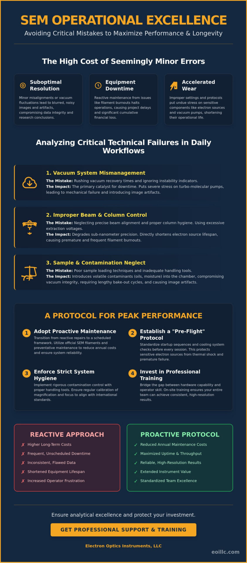
Incorrect beam settings represent one of the most frequent causes of accelerated cathode degradation. When an operator applies excessive extraction voltages or ignores saturation levels, they directly shorten the lifespan of the electron source, leading to premature filament burnout. Furthermore, poor sample loading techniques often introduce volatile contaminants into the chamber. These contaminants compromise the vacuum’s integrity and require lengthy bake-out cycles to rectify. It’s critical to respect vacuum recovery times; rushing the transition from atmosphere to high vacuum puts undue stress on the turbo-molecular pumps, potentially leading to mechanical failures that necessitate professional repair visits.
Data Integrity and the Cost of Image Artifacts
Operational neglect doesn’t just damage hardware; it compromises the very data the laboratory exists to produce. Image artifacts, such as charging, drift, or uncorrected astigmatism, often lead to false data interpretation and flawed conclusions. If an initial imaging session yields poor results due to user error, the cost of repeating that research—including labor, consumables, and instrument time—can be staggering. Establishing a baseline for acceptable imaging performance involves several key steps:
- Regular Calibration: Ensuring magnification and focus scales align with international standards.
- Beam Alignment: Minimizing aperture errors to ensure the electron probe is perfectly centered.
- Contamination Control: Utilizing proper handling tools to prevent oil or moisture from entering the column.
By avoiding common sem operational mistakes, research teams can ensure that every scan contributes to a valid, high-resolution dataset that stands up to peer review and industrial scrutiny.
Analyzing the Critical Technical Failures in Daily SEM Workflows
Daily workflows in a high-precision laboratory environment demand more than mere software proficiency. They require a rigorous comprehension of the physical interactions occurring within the electron column. Identifying common sem operational mistakes often begins with recognizing that the instrument is a dynamic system, sensitive to the slightest deviations in its internal environment. While modern hardware provides significant automation, the operator’s ability to interpret system feedback remains the cornerstone of consistent, high-resolution results.
Improper vacuum system management stands as the primary cause of scanning electron microscopy downtime. A vacuum is not a static state but a delicate equilibrium that can be easily disrupted by improper handling or the introduction of unsuitable materials. When vacuum integrity is compromised, the mean free path of electrons is reduced, leading to increased scattering and a noticeable degradation in image contrast. Understanding these fundamental SEM operational principles is essential for diagnosing why a system fails to reach its specified resolution or why filaments expire prematurely.
Vacuum Management and Contamination Control
The introduction of outgassing materials into the chamber is a frequent error that leads to long-term column contamination. Porous samples, oily residues, or even unvented mounting adhesives release volatile molecules that migrate toward the electron source and the final aperture. To maintain the integrity of the system, operators must practice meticulous hygiene, including the use of powder-free gloves and the proper lubrication of O-rings and door seals. Early signs of pump inefficiency, such as erratic vacuum recovery times or unusual acoustic signatures from the turbo pump, should prompt an immediate review of operational protocols before a full system shutdown occurs. Regular preventative maintenance visits can help identify these mechanical stressors before they result in hardware failure.
Mastering Beam Alignment and Astigmatism Correction
Beam alignment is not a one-time setup procedure but a necessary daily adjustment. Thermal expansion of column components and minor magnetic drift mean that the electron probe’s path must be periodically centered to ensure peak spatial resolution. Failure to perform these alignments results in “comatic” aberration, where the beam shape becomes distorted, leading to blurred edges and lost detail. Correcting astigmatism is equally critical; operators should utilize high-magnification features to adjust the stigmator coils until the image remains sharp across both the X and Y axes. A common mistake involves choosing an excessively high accelerating voltage for sensitive samples, which causes subsurface beam penetration and masks the surface topography you intend to capture.
Sample Preparation: The Overlooked Operational Step
Data quality is inextricably linked to the physical stability of the specimen. Poor mounting techniques lead to mechanical instability and image drift, particularly at high magnifications where even nanometer-scale movements are visible. For non-conductive specimens, the necessity of a uniform conductive coating cannot be overstated. Without it, the specimen will accumulate negative charge, creating a localized electric field that deflects the primary beam and produces “blooming” artifacts. Mastering these nuances of sem operation ensures that your research is defined by clarity rather than by unforced technical errors. Neglecting environmental factors like electromagnetic interference or floor vibrations can also undermine your work, making it vital to monitor the lab’s physical surroundings during sensitive imaging sessions.
Addressing the Misconception of Equipment Age vs. Maintenance Neglect
Many laboratory directors mistakenly attribute declining image quality to the chronological age of their instrumentation. In reality, a well-maintained system from a previous decade can often outperform a newer model that has been subjected to common sem operational mistakes. The distinction between inevitable hardware wear and avoidable user-induced damage is critical for optimizing long-term capital investments. While mechanical components like vacuum seals and stage motors possess finite lifecycles, their degradation is frequently accelerated by poor operational hygiene rather than mere time. A disciplined approach to hardware stewardship ensures that technical authority remains grounded in consistent, high-resolution output.
The “it still works” mentality represents a significant risk to system integrity. Using consumable parts beyond their recommended operational hours doesn’t just reduce image quality; it risks secondary damage to the electron column. For instance, a thinning filament might continue to emit electrons but can eventually snap, causing a potential surge that damages the high-voltage supply or contaminating the gun chamber with debris. Integrating a rigorous sem maintenance strategy ensures that these risks are mitigated through proactive replacement rather than reactive repair. This transition from a reactive to a visionary maintenance framework is what separates world-class research facilities from those struggling with inconsistent data.
The Lifecycle of SEM Filaments and Apertures
Apertures and filaments are the essential consumables of the SEM; they are designed for periodic replacement to maintain beam stability. Tungsten filaments are valued for their robustness, yet they require precise saturation to avoid premature thinning. Conversely, Cerium Hexaboride (CeB6) cathodes offer higher brightness and extended life but remain significantly more sensitive to vacuum fluctuations. Contaminated apertures often manifest as uncorrectable astigmatism or a noticeable loss of beam current stability. If you observe a “comet tail” effect on high-contrast edges that cannot be resolved through standard alignment, it’s an empirical sign that the final aperture requires professional cleaning or replacement to restore imaging precision.
Preventative Maintenance: A Strategic Investment
Annual service contracts and scheduled preventative maintenance visits shouldn’t be viewed as discretionary expenses but as strategic safeguards for laboratory uptime. Professional calibration of electromagnetic lenses and secondary electron detectors ensures that the instrument’s output remains accurate to its original factory specifications. Establishing a comprehensive logbook allows teams to track performance trends, such as vacuum pump-down times and filament hours. This data-driven approach enables the identification of subtle system drifts before they manifest as critical failures. By documenting these variables, organizations can maximize the return on investment and ensure their hardware continues to push the boundaries of what is possible in advanced materials analysis.
Establishing a Protocol for Peak Performance: The SEM Operational Checklist
The transition from theoretical knowledge to operational excellence requires a structured framework that mitigates the risk of human error. Implementation of a rigorous daily checklist is the most effective defense against common sem operational mistakes that compromise both data integrity and hardware longevity. This protocol ensures that every variable, from vacuum stability to detector optimization, is verified before the electron beam ever interacts with the specimen. By standardizing these pre-flight checks, laboratories can achieve a level of systemic stability that mirrors the reliability found in high-end industrial production environments.
A visionary approach to microscopy involves recognizing that the instrument’s performance is a reflection of its environment. Daily pre-flight checks must include the verification of cooling systems, as even minor fluctuations in chiller temperature can induce thermal drift in the electromagnetic lenses. Similarly, monitoring gas pressures for variable pressure modes and ensuring vacuum levels reach their specified base pressure are non-negotiable steps. Standardizing the startup sequence acts as a primary safeguard for the electron source, ensuring that the filament is saturated gradually to prevent thermal shock. This meticulousness is what allows advanced hardware, such as the Veritas Series SEM, to maintain its sub-nanometer resolution over extended research cycles.
The Daily Startup and Calibration Routine
A professional startup sequence follows a logical progression designed to protect the column’s internal components. The following steps should be performed at the commencement of every session:
- Step 1: Verifying Vacuum Stability: Confirm that the chamber pressure has reached its optimal high-vacuum state and that the ion pump readings are consistent with historical performance data.
- Step 2: Filament Saturation and Centering: Increase the heating current methodically until the first saturation point is reached, then center the filament within the grid cap to ensure a symmetrical beam profile.
- Step 3: Resolution Testing: Utilize a standard calibration sample, such as gold-on-carbon, to verify that the system achieves its rated resolution and that there is no evidence of uncorrected astigmatism.
Optimizing Imaging Parameters for High-Resolution Results
Selecting the correct imaging parameters is a strategic decision that depends on the specific goals of the analysis. For surface topography, a short working distance (WD) is often preferred to minimize the effects of lens aberrations, while a longer WD provides the necessary depth of field for complex, three-dimensional structures. The choice of spot size involves a similar trade-off; smaller spot sizes are essential for high-resolution imaging, whereas larger spot sizes provide the higher probe current needed for compositional analysis via backscattered electron (BSE) detectors. The magnitude of the probe current serves as the primary determinant for image noise levels, necessitating a precise balance to maintain edge definition while ensuring sufficient signal for detector acquisition.
To ensure your laboratory consistently achieves these superior standards, it’s essential to integrate these technical SEM operation protocols into your team’s daily workflow. Following a disciplined post-operation shutdown—including venting with dry nitrogen and ensuring the column is isolated—will further preserve the internal vacuum integrity for future use.
Ensuring Analytical Excellence Through Professional Support and Training
While previous sections detailed the technical mechanics of vacuum stability and beam alignment, the human element remains the final frontier in achieving sub-nanometer precision. To better understand how technicians interact with complex imaging systems, labs can utilize behavioral research software from Ergoneers to identify patterns that lead to operational errors. Analytical excellence isn’t merely a byproduct of superior hardware; it’s cultivated through a rigorous commitment to professional development and on-site technical training. By refining the operator’s understanding of electron optics, laboratories can effectively eliminate the common sem operational mistakes that often stem from a surface-level engagement with the instrument’s interface.
Customized Training for Specialized Applications
Generic manuals often lack the depth required for complex failure analysis or specialized materials characterization. Manufacturer-direct training on specific platforms, such as the Cube II Benchtop SEM or the Genesis Tabletop SEM, provides laboratory staff with the nuanced insights necessary to push these systems to their physical limits. This specialized education reduces operator-induced variability, ensuring that high-stakes research remains reproducible across different shifts and team members. Integrating advanced sem techniques into the standard lab workflow allows for more sophisticated data acquisition, such as low-voltage imaging of beam-sensitive polymers or high-speed EDS mapping that captures compositional data without compromising spatial resolution.
The physical integrity of an SEM is most vulnerable during periods of transition, such as installation or relocation. Hidden operational flaws often originate from improper column leveling, inadequate vibration isolation, or the subtle misalignment of electromagnetic shielding during the initial setup. Professional installation ensures that the vacuum system and electron optics are calibrated to their specific environmental conditions, preventing the long-term drift and instability that can plague improperly handled systems. Engaging expert service for these complex tasks ensures that the system avoids the common sem operational mistakes associated with amateur hardware handling, protecting your investment from the moment of commissioning.
Partnering for Long-Term Laboratory Success
Partnering with an established high-tech authority like Electron Optics Instruments, LLC provides laboratories with a national support network that’s essential for maintaining uptime in critical industrial sectors. Comprehensive service contracts offer priority access to critical components, including high-brightness filaments and EDS detectors, while ensuring that preventative maintenance is performed by seasoned engineers who understand the intersection of innovation and reliability. This partnership reflects the Electron Optics Instruments, LLC commitment to 30 years of electron microscopy excellence, providing a stable foundation for global innovators to pursue the next generation of hardware and technical systems. By leveraging this external expertise, your laboratory can transition from basic operation to a visionary research model that consistently delivers superior standards of accuracy.
Advancing Research Standards through Operational Excellence
Operational excellence in scanning electron microscopy is not a destination but a continuous commitment to technical meticulousness. By systematically eliminating common sem operational mistakes, your laboratory can transform its imaging capabilities from inconsistent output to a steady stream of high-resolution data. We’ve explored how vacuum integrity, precise beam alignment, and rigorous maintenance protocols act as the cornerstones of hardware longevity and analytical accuracy. These practices ensure that sophisticated instruments, like the Veritas FE SEM or the Cube II Benchtop SEM, continue to operate at the pinnacle of their design specifications.
Achieving this level of systemic reliability requires a partner that understands the intersection of innovation and practical production. With over 30 years of industry experience, Electron Optics Instruments, LLC serves as the sole US distributor for EmCraft SEMs and provides comprehensive service for all major SEM manufacturers. Whether you require preventative maintenance visits or specialized on-site training, our expertise ensures your facility maintains its competitive edge in a rapidly evolving industrial landscape. Contact Electron Optics Instruments, LLC for Expert SEM Service and Training to secure the future of your research mission.
Frequently Asked Questions
What are the most common causes of poor SEM image quality?
Suboptimal image resolution is typically the result of uncorrected astigmatism, improper beam alignment, or inadequate vacuum levels. These common sem operational mistakes prevent the electron probe from achieving its theoretical spot size, leading to blurred edges and a significant loss of topographic detail. Environmental factors, such as electromagnetic interference or floor vibrations, can also introduce periodic noise that compromises the analytical integrity of high-magnification scans.
How often should an SEM vacuum system be serviced?
A comprehensive service of the vacuum system should be conducted annually during scheduled preventative maintenance visits to ensure systemic stability. This interval allows for the inspection of turbo-molecular pumps, the replacement of rotary pump oil, and the verification of O-ring integrity. Regular servicing prevents the accumulation of contaminants that can lead to unexpected vacuum failures and prolonged instrument downtime in high-stakes industrial environments.
Can improper sample preparation damage the SEM column?
Yes, introducing inadequately dried or contaminated specimens can cause volatile molecules to outgas and migrate throughout the electron column. These contaminants often settle on sensitive components like the final aperture or the electron source, leading to beam instability and permanent hardware degradation. Meticulous sample preparation is a fundamental requirement for protecting the sophisticated optics found in instruments like the Veritas Series SEM.
What is the difference between astigmatism and poor focus in SEM?
Focus refers to the convergence of the electron beam at the precise plane of the specimen surface, whereas astigmatism is a geometric distortion of the beam’s cross-section. While a poorly focused image appears uniformly soft, astigmatism causes the image to “stretch” or blur along different axes as the focus is adjusted. Correcting this distortion requires the precise manipulation of stigmator coils to restore a perfectly circular beam profile.
Why does my SEM filament keep burning out prematurely?
Premature filament failure is almost always caused by operating the cathode at excessive heating currents or within a compromised vacuum environment. When an operator bypasses the recommended saturation point, the tungsten or CeB6 material thins rapidly due to evaporation, significantly shortening its functional lifecycle. Adhering to standardized startup sequences and monitoring filament hours are critical steps in reducing annual consumable costs.
Is on-site SEM training necessary for experienced lab technicians?
On-site training is essential because the software architectures and hardware nuances of modern systems, such as the Cube II Benchtop SEM, differ significantly across manufacturers. Professional training ensures that even seasoned technicians can master specific advanced hardware features and avoid common sem operational mistakes unique to a new installation. This specialized education bridges the gap between general microscopy knowledge and the high-end precision required for specialized industrial applications.
How do environmental vibrations affect benchtop SEM performance?
Environmental vibrations introduce mechanical instability that manifests as jagged edges or “sawtooth” artifacts in high-magnification images. Even compact systems like the Genesis Tabletop SEM are sensitive to low-frequency oscillations from nearby heavy machinery or HVAC systems. Proper site selection and the use of specialized vibration isolation platforms are necessary to ensure that these external variables don’t undermine the system’s sub-nanometer imaging capabilities.
What should I do if my SEM vacuum fails to reach the base pressure?
You should immediately inspect the chamber door seals and O-rings for microscopic debris or hair that may be preventing a hermetic seal. If the vacuum levels don’t improve after a thorough cleaning of the access points, it’s necessary to check the performance of the pre-vacuum pumps and verify that there are no leaks in the gas injection lines. Persistent vacuum issues require professional technical intervention to prevent damage to the turbo pump or the electron gun chamber.