Could a microscopic layer of carbonized residue on a platinum disc be the sole barrier between your laboratory and sub-nanometer clarity? For many professionals operating high-end instrumentation in 2026, the frustration of persistent image blurring and stubborn astigmatism often persists despite exhaustive alignment efforts. It’s a common struggle to balance the requirement for pristine optics with the inherent risks of handling delicate components, especially when the spot price of platinum has reached approximately $1,890.40 per ounce. Implementing a standardized sem aperture cleaning procedure is no longer a luxury but a fundamental necessity for maintaining the technical integrity of your analytical results.
This guide outlines the rigorous protocols required to eliminate contamination and restore the diffraction-limited performance of your scanning electron microscope. You’ll master the delicate balance of manual decontamination using 1-micron diamond polishing paste and learn to leverage modern software environments, such as ZEISS ZEN core 3.13, to monitor system health. We provide a comprehensive overview of safe maintenance practices, ranging from thermal evaporation techniques to the latest in-situ cleaning technologies, ensuring your team can extend the operational lifespan of your apertures while achieving the consistent, high-resolution imaging that defines professional excellence.
Key Takeaways
- Recognize the specific pathways through which hydrocarbon thin-films accumulate during beam-sample interactions to proactively mitigate image degradation.
- Execute a standardized sem aperture cleaning procedure that integrates chemical, thermal, or plasma-based modalities to restore sub-micron resolution and analytical accuracy.
- Establish a controlled environment for manual decontamination protocols, utilizing specialized instrumentation like non-magnetic tweezers and ultrasonic baths to maintain component integrity.
- Diagnose post-procedural imaging anomalies by evaluating the influence of residual solvent films on vacuum levels and ensuring precise mechanical repositioning of the aperture strip.
- Implement long-term operational strategies that combine scheduled preventative maintenance with the utilization of advanced benchtop systems, such as the Cube II, to optimize lab productivity.
Understanding the Impact of Aperture Contamination on SEM Performance
The objective aperture serves as a critical spatial filter within the Scanning Electron Microscope (SEM), functioning to define the final probe diameter by excluding peripheral electrons that contribute to spherical aberration. By refining the convergence angle of the electron beam, this small platinum or molybdenum disc ensures that the probe remains coherent and tightly focused upon the sample surface. Over time, however, the purity of this component is compromised by the accumulation of hydrocarbon thin-films. These contaminants are typically the result of beam-induced polymerization of residual organic molecules within the vacuum chamber, often sourced from sample outgassing or vacuum system lubricants. As these layers thicken, they transform the aperture from a passive geometric constraint into an active source of imaging instability.
This non-conductive residue leads to a phenomenon known as “charging,” where incident electrons accumulate on the contamination layer rather than being safely shunted to ground through the metal disc. This buildup creates a localized electrostatic field that interacts with the primary beam. The primary symptoms of this degradation include intractable astigmatism that defies standard stigmator correction, noticeable beam drift during high-resolution scans, and a significant loss of edge definition. Without a rigorous sem aperture cleaning procedure, these issues eventually render sub-micron analysis impossible and can lead to inconsistent data across long-term research projects.
The Physics of Beam Deflection and Image Aberration
When a non-uniform charge distribution develops on the aperture’s edge, it functions as a parasitic lens. This unwanted electrostatic field exerts a transverse force on the electron beam, causing spot shape asymmetry where the probe becomes elongated rather than perfectly circular. At higher magnification settings, the impact is magnified. The smaller scan area requires a more stable probe, yet the relative influence of the parasitic lens becomes more dominant, leading to a visible “smearing” of topographical features. This distortion is particularly problematic in 2026 as laboratories push for higher resolution at lower accelerating voltages, where the beam is more susceptible to external fields.
Distinguishing Between Condenser and Objective Aperture Issues
Differentiating between contamination points is vital for efficient maintenance. Condenser apertures, situated higher in the column, typically experience higher contamination rates due to direct exposure to the high-intensity source beam, manifesting as erratic beam current fluctuations or “flicker” in the secondary electron signal. In contrast, fouling of the final objective aperture directly degrades spatial resolution. Technicians often utilize the “wobbler” function to diagnose these shifts; if the image exhibits lateral translation instead of a symmetric expansion and contraction about the optical axis, it’s a definitive indicator that the objective aperture requires a formal sem aperture cleaning procedure to restore optical alignment.
Technical Modalities for SEM Aperture Cleaning
The selection of an appropriate decontamination modality represents a strategic intersection between chemical efficacy and material preservation. In the context of 2026 laboratory standards, the choice of a specific sem aperture cleaning procedure must be dictated by the material composition of the disc and the specific density of the accumulated foulants. Platinum remains the preferred material for high-performance imaging due to its superior electron-stopping power, yet its current market valuation of approximately $1,890.40 per ounce demands a protocol that prioritizes component longevity. Molybdenum and gold foil apertures offer alternative conductive profiles; each requires a distinct technical approach to ensure that the cleaning process does not introduce structural deformities or alter the critical dimensions of the aperture’s micro-geometry.
Chemical and Solvent-Based Decontamination
High-purity acetone and isopropanol serve as the primary agents for removing non-polymerized oils and loose particulates during the initial stages of maintenance. While these solvents are indispensable for degreasing, they lack the reactive energy to penetrate the dense, cross-linked carbon structures formed during prolonged high-voltage beam exposure. Users should exercise extreme caution regarding drying protocols. Improper evaporation can lead to the entrapment of solvent molecules within the aperture’s orifice, subsequently causing vacuum instability or secondary contamination upon beam re-initiation. This method is best reserved for routine, low-density cleaning cycles where the risk of heavy carbonization is minimal.
Thermal Evaporation and Flaming Procedures
For more resilient carbonaceous deposits that resist chemical dissolution, thermal evaporation remains a reliable industrial standard. This process involves utilizing a specialized aperture flamer or a vacuum furnace to raise the temperature of the component until hydrocarbons are vaporized. Molybdenum apertures are particularly well-suited for this high-heat treatment due to their exceptional thermal stability and high melting point. While platinum apertures also tolerate flaming, the operator must maintain precise control over the temperature to prevent mechanical warping or the recrystallization of the metal. Such structural changes could compromise the circularity of the aperture hole, leading to permanent aberrations in the electron probe.
Plasma Discharge and In-Situ Cleaning
The contemporary shift toward plasma-based decontamination reflects a broader industry trend toward non-destructive, high-precision maintenance systems. By utilizing a controlled discharge of oxygen or argon plasma, the system induces a chemical reaction that oxidizes carbon contaminants into volatile gases, which are then evacuated by the vacuum system. This “gentle” approach is increasingly integrated into modern architectures like the Cube II benchtop system. Adopting these professional SEM consumables and maintenance strategies allows labs to perform in-situ cleaning, significantly reducing the necessity for frequent column disassembly and minimizing the risk of mechanical damage. Ultimately, selecting a sophisticated sem aperture cleaning procedure ensures that the synergy between hardware reliability and imaging precision remains uninterrupted.

Step-by-Step Manual Aperture Cleaning Procedure
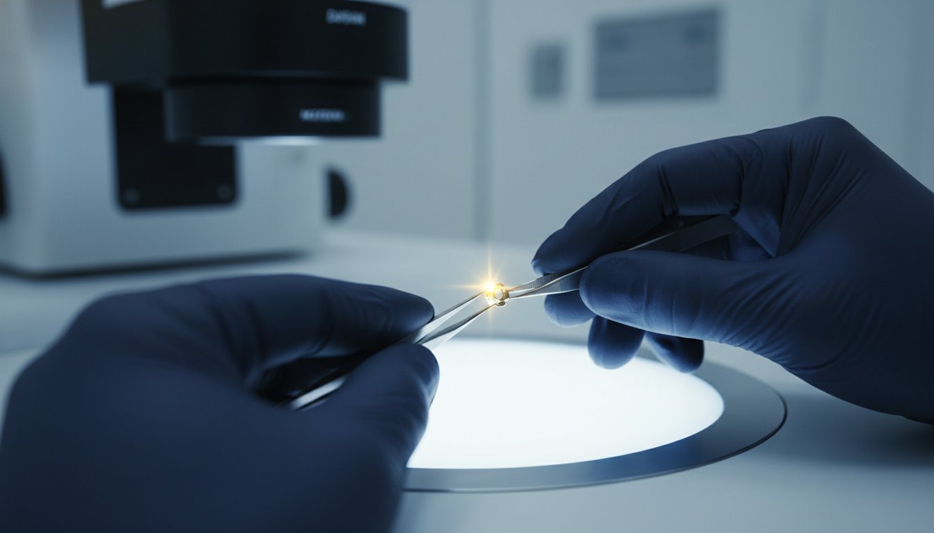
Executing a manual sem aperture cleaning procedure necessitates a strictly controlled environment to mitigate the risk of introducing ambient particulates into the sensitive electron column. Technicians should ideally perform these operations within a Class 100 clean-room or under a dedicated laminar flow hood to ensure that the cleaning process doesn’t inadvertently become a source of new contamination. Maintaining the structural integrity of these micro-scale components requires a suite of specialized tools, including high-precision non-magnetic tweezers, a multi-stage ultrasonic bath, and lint-free, clean-room grade wipes. Using titanium or carbon-tipped tweezers is essential, as standard steel tools can introduce magnetic interference or cause micro-scratches on the delicate platinum or molybdenum surfaces.
Stage 1: Extraction and Initial Inspection
The process begins with the systematic venting of the SEM column according to the specific vacuum protocols of your instrument, ensuring the chamber reaches atmospheric pressure before the aperture holder is retracted. Once the assembly is removed, a rigorous visual assessment is vital to determine the severity of fouling. It’s important to distinguish between reversible hydrocarbon buildup and irreversible physical damage, such as pitting or electron-beam-induced melting of the aperture edge. The gold standard for preliminary aperture inspection involves evaluating the orifice under 40x optical magnification to confirm the presence of concentric contamination rings or structural defects.
Stage 2: Ultrasonic Cleaning and Solvent Rinsing
Following the initial inspection, the aperture undergoes a sequential chemical bath to dissolve polymerized layers. It’s best to initiate the process with a high-purity acetone immersion in the ultrasonic bath for approximately three minutes to degrease the surface. This is followed by a secondary rinse in electronic-grade ethanol to eliminate any residual acetone films. Operators must monitor ultrasonic duration closely, as excessive exposure can induce mechanical stress or fatigue in thin metal foils. Final drying is achieved using a steady, low-pressure stream of ultra-pure compressed nitrogen, which ensures that no solvent molecules remain trapped within the micro-aperture to outgas during subsequent vacuum cycles.
Stage 3: Reinstallation and Alignment
Reintegrating the cleaned component requires meticulous handling to preserve vacuum integrity and avoid the introduction of skin oils. After reseating the aperture strip into its holder, the assembly is reinserted, and the system is pumped down to its operational vacuum level. Precise mechanical alignment is mandatory following any manual intervention, as even a micron-scale shift can deviate the beam from the optical axis. After the vacuum stabilizes, the operator should perform a full beam-centering routine to restore imaging precision. For comprehensive guidance on maintaining high-performance instrumentation, technicians should adhere to established SEM operation best practices to ensure long-term system reliability.
Troubleshooting Post-Cleaning Imaging Anomalies
The completion of a rigorous sem aperture cleaning procedure does not always result in immediate imaging restoration, often leading to what technicians describe as the ‘Clean but Blurry’ paradox. This phenomenon is frequently the result of subtle mechanical misalignment during the re-insertion of the aperture strip. While the component may be chemically pristine, a lateral shift of even a few microns can deviate the beam from the optical axis, introducing severe off-axis aberrations. Additionally, laboratories must account for the influence of residual solvent films. If the drying phase using high-purity nitrogen was insufficient, trapped acetone or ethanol molecules will outgas, causing localized vacuum fluctuations that manifest as beam instability or image flicker.
To ensure a successful return to operation, technical teams should utilize a standardized checklist for verifying column vacuum recovery and imaging stability:
- Confirm that the ion pump or Penning gauge returns to baseline vacuum levels within 10 to 15 minutes of re-insertion.
- Monitor the emission stability; erratic beam current often indicates residual outgassing near the final lens assembly.
- Perform a crossover check to verify that the beam remains centered as the condenser lens strength is modulated.
If the central orifice appears non-circular or exhibits jagged edges under optical inspection, the aperture is considered ‘blown.’ Such structural damage is typically caused by accidental beam strikes during high-current operations or the use of incorrect accelerating voltages. Unlike hydrocarbon buildup, this physical pitting cannot be rectified through standard cleaning modalities and requires immediate component replacement to restore the electron probe’s symmetry.
Persistent Astigmatism and Beam Instability
When astigmatism remains intractable after a confirmed sem aperture cleaning procedure, the contamination source likely resides further up the column. Hydrocarbon buildup on the column liners or within the electron gun area can create parasitic electrostatic fields that mimic the symptoms of aperture charging. Technicians can distinguish between these by observing the frequency of the instability. Electrical noise typically presents as high-frequency jitter, whereas aperture charging manifests as a slow, rhythmic drift or a sudden jump in the image. In cases of systemic column fouling, a full high-temperature bake-out is often more effective than repeated aperture maintenance.
Evaluating Aperture Lifespan and Replacement Triggers
There are definitive physical limits to the efficacy of manual decontamination. Once a disc has undergone multiple thermal flaming cycles or shows signs of thinning at the orifice edge, it is considered beyond repair. Molybdenum apertures, while cost-effective, are more prone to oxidative stress than platinum, making frequent replacement a more viable strategy than repeated cleaning. Maintaining a robust inventory of high-quality SEM consumables ensures that lab operations are not stalled by component fatigue. For labs seeking to minimize downtime and maintain international standards of accuracy, investing in professional SEM filaments and consumables provides the necessary reliability for continuous, high-precision analytical workflows.
Professional Maintenance Strategies for Industrial SEM Systems
The strategic integration of a systematic sem aperture cleaning procedure into a broader laboratory workflow is essential for maintaining operational continuity in high-throughput industrial environments. While individual technicians can master manual decontamination, the long-term reliability of a scanning electron microscope relies on a holistic maintenance framework that balances internal protocols with professional oversight. This dual-track approach minimizes the risk of unexpected downtime and ensures that imaging precision remains within the specified tolerances for sub-micron analysis. It’s a strategy that reflects a commitment to both industrial performance and the meticulousness required for international production excellence, ensuring that the intersection of innovation and reliability remains undisturbed.
Maximizing Uptime with Benchtop SEM Solutions
The evolution of benchtop technology has significantly lowered the barrier to entry for routine maintenance without compromising analytical depth. The Cube II Benchtop SEM, for instance, features a modular column architecture that facilitates more direct access to the aperture assembly compared to traditional floor-standing units. This design philosophy allows operators to perform a sem aperture cleaning procedure with greater confidence, and it’s a significant factor in reducing the risk of mechanical misalignment. When these systems are equipped with EDS (Energy Dispersive Spectroscopy) Systems, the cleanliness of the aperture becomes even more critical. Hydrocarbon buildup can produce stray electron scattering that degrades the signal-to-noise ratio of elemental maps, leading to inconsistent data. On-site technical training programs further empower lab teams by standardizing these cleaning techniques, ensuring that every operator adheres to the same rigorous benchmarks for cleanliness and alignment.
The Role of Annual Preventative Maintenance
Professional service visits represent a critical anchor for industrial labs, providing a level of technical depth that exceeds routine user intervention. Managing complex electron optics doesn’t have to be a source of operational anxiety when a structured protocol is in place. An annual preventative maintenance visit includes a comprehensive assessment of the entire electron optical column, involving the deep cleaning of gun components and the precise calibration of electromagnetic lenses. This calibration is vital; it ensures that the magnetic lens fields remain perfectly symmetric, which is a prerequisite for the aperture to function as an effective spatial filter. By aligning these complex hardware systems, EOI helps labs maintain a steady, expert-driven narrative of success and technical prowess. Explore our comprehensive SEM service contracts to ensure peak performance and secure a visionary partner for your technical training and complex repair requirements.
Advancing Analytical Accuracy Through Rigorous Maintenance Standards
Maintaining the integrity of sub-micron imaging requires a proactive stance against the inevitable accumulation of hydrocarbon contaminants. By implementing a standardized sem aperture cleaning procedure, laboratories ensure that their instrumentation remains a reliable vehicle for innovation rather than a source of analytical frustration. You’ve explored how identifying specific fouling markers and utilizing appropriate chemical or plasma-based modalities can restore the diffraction-limited performance of your scanning electron microscope. These technical protocols represent the baseline for professional excellence in 2026.
When internal maintenance reaches its limits or when complex system calibrations are required, partnering with an established industry authority becomes essential. Electron Optics Instruments brings over 30 years of electron microscopy expertise to your facility, serving as the sole US distributor for EmCraft SEMs and providing authorized service for all major SEM manufacturers. We’re dedicated to ensuring your research and production environments achieve peak performance through meticulous hardware support. Contact Electron Optics Instruments for Expert SEM Maintenance and Service to secure the future of your imaging precision. Your commitment to technical accuracy is the catalyst for the next generation of industrial breakthroughs.
Frequently Asked Questions
How often should SEM apertures be cleaned in a high-use lab?
In high-throughput industrial environments, objective apertures typically require inspection every three to six months. If your instrument is utilized for high-current applications or analyzes outgassing samples, this frequency may increase to a monthly schedule. Monitoring the stigmator values provides a reliable indicator; when the correction required to achieve a circular probe exceeds established baselines, it’s time to initiate maintenance.
Can I use standard laboratory-grade solvents for aperture cleaning?
You must utilize ultra-pure, electronic-grade solvents like acetone and ethanol to prevent the introduction of non-volatile residues. Standard laboratory-grade solvents often contain trace impurities that leave a thin film upon evaporation. These films subsequently outgas under high vacuum, leading to beam instability and secondary contamination of the column liners, which compromises imaging precision.
What is the difference between platinum and molybdenum apertures regarding maintenance?
Platinum apertures offer superior chemical resistance and can withstand repeated thermal flaming, making them ideal for long-term use despite the spot price of platinum being approximately $1,890.40 per ounce. Molybdenum alternatives are more susceptible to oxidative stress and mechanical warping at high temperatures. While molybdenum is cost-effective for high-turnover labs, it generally requires more frequent replacement cycles to maintain structural integrity.
Is it possible to clean a self-cleaning gold foil aperture?
Self-cleaning gold foil apertures are designed to utilize beam-induced heating to vaporize contaminants, but they aren’t immune to permanent carbonization. Manual cleaning is generally discouraged due to the extreme fragility of the foil. If the “self-cleaning” mechanism fails to maintain image clarity, the industry standard is to replace the disc rather than risk mechanical destruction through manual scrubbing or ultrasonic treatment.
What are the risks of using an ultrasonic cleaner on thin SEM apertures?
The primary risk of ultrasonic cleaning is mechanical fatigue or structural cracking of the thin metal disc. High-frequency vibrations can induce micro-fractures, especially in apertures with high aspect-ratio orifices or those made of gold foil. To mitigate this risk, you should limit the immersion time to approximately three to five minutes and ensure the component is properly supported in a specialized holder.
How can I tell if my imaging issues are caused by the aperture or the filament?
Filament degradation typically manifests as erratic emission current or a total loss of beam saturation, whereas aperture issues cause localized image aberrations. If you observe directional blurring or a loss of edge definition that persists after stigmator adjustment, the aperture is the likely culprit. Filament health is better diagnosed through the emission monitoring tools in modern control software, which can track source age and stability.
What safety precautions are necessary when handling SEM apertures?
Handling these micro-components requires the use of non-magnetic, titanium-tipped tweezers and powder-free nitrile gloves to prevent the transfer of skin oils. All procedures should be conducted within a laminar flow hood to avoid ambient dust. Because these discs are easily lost or damaged, maintaining a dedicated, organized workstation is a fundamental safety requirement for the sem aperture cleaning procedure.
Should I clean or replace my apertures if I see beam drift?
You should always attempt a standardized sem aperture cleaning procedure before opting for replacement, provided there’s no visible physical damage. If the beam drift or astigmatism persists after thorough decontamination and a full column alignment, the aperture has likely reached its physical lifespan. In such cases, replacing the component is the only way to restore the sub-micron resolution required for advanced hardware analysis.