The traditional reliance on optical light microscopy for micro-trace evidence is no longer sufficient to meet the rigorous standards of 2026 legal scrutiny. You likely recognize that the inherent inability to distinguish between chemically similar yet morphologically distinct materials can compromise the integrity of an entire investigation, especially when light-based systems reach their physical limits. The persistent pressure to process high volumes of evidence while maintaining meticulous standards of legal defensibility remains a primary concern for practitioners who require absolute certainty in their analytical outcomes. This article demonstrates how advanced sem for forensic investigation provides the definitive morphological and elemental evidence required for high-stakes cases. By utilizing sophisticated systems like the Veritas Series or Genesis Tabletop SEM, professionals can achieve magnifications exceeding 100,000x and resolutions below 1 nm. We’ll examine how streamlined, automated workflows and adherence to new standards such as ANSI/ASTM E3497-26 facilitate the unambiguous identification of gunshot residue and other trace materials. You’ll learn how the integration of Energy Dispersive Spectroscopy (EDS) ensures that every data point remains legally defensible and scientifically robust.
Key Takeaways
- Understand why modern forensic standards require sub-micron topographical mapping that exceeds the physical resolution limits of traditional optical systems.
- Learn how integrating EDS systems with sem for forensic investigation provides a definitive elemental fingerprint for gunshot residue and complex trace materials.
- Explore the automated workflows designed to identify the specific lead-barium-antimony signatures required for legally defensible evidence.
- Evaluate the strategic shift toward benchtop solutions like the Cube II and Genesis to facilitate rapid-response forensic analysis without sacrificing data integrity.
- Discover how the Veritas Series maintains high-volume evidence processing while ensuring a rigorous chain of custody for all digital analytical data.
The Evolution of Forensic Microscopy: Why SEM is Essential in 2026
Forensic science has transitioned into an era where the most critical evidence exists beyond the threshold of human vision, necessitating a shift toward instruments that provide both morphological and chemical certainty. Traditional optical systems, while useful for macroscopic survey, lack the necessary depth of field and lateral resolution required to characterize the complex particulates found at modern crime scenes. Adopting sem for forensic investigation represents a strategic evolution in laboratory capability, allowing technicians to move beyond simple magnification toward comprehensive topographical and elemental characterization. This shift from analog observation to digital, high-fidelity electron-based data ensures that every micro-trace, from a single fiber to a microscopic paint chip, is documented with a level of precision that satisfies the most rigorous international standards.
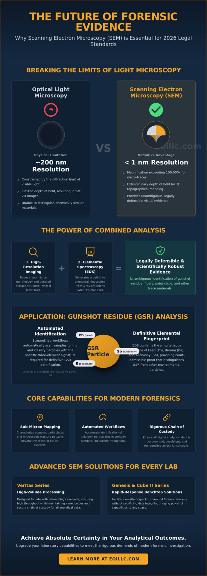
Breaking the Limits of Light Microscopy
The physical constraints of visible light create a definitive barrier known as the diffraction limit, which fundamentally restricts the clarity of images at high magnifications. Because the wavelengths of light are significantly longer than those of accelerated electrons, optical microscopes cannot resolve features smaller than half the wavelength of the light used, rendering them ineffective for sub-micron analysis. In contrast, the Scanning electron microscope (SEM) utilizes a focused beam of electrons to bypass these limitations, providing an extraordinary depth of field that renders samples in three dimensions. This topographical mapping is vital for ballistics and tool mark analysis, where identifying microscopic fracture patterns or striations requires more than just a flat, magnified image. While optical microscopes are physically constrained by the diffraction limit of light to approximately 200 nanometers, a scanning electron microscope (SEM) achieves resolutions of less than 1 nanometer, enabling the visualization of features thousands of times smaller.
The Role of High-Fidelity Imaging in Legal Admissibility
Legal defensibility in 2026 rests upon the systematic removal of subjective interpretation from the evidentiary chain, a goal achieved through the deployment of advanced analytical hardware. High-resolution imaging provides an empirical foundation that strengthens the weight of evidence presented to a jury, as the clarity of a sem scanning electron microscope image leaves little room for the ambiguity often associated with traditional methods. By standardizing digital imaging protocols for national forensic databases, laboratories ensure that data remains consistent and reproducible across various international jurisdictions. This transition to high-fidelity imaging doesn’t just improve visual clarity; it significantly accelerates the identification of unknown particulates in complex samples, allowing investigators to isolate and analyze individual grains of sand or shards of glass that would be indistinguishable under light-based systems. It’s this commitment to technical accuracy and meticulousness that defines the modern forensic workflow, ensuring that the intersection of innovation and reliability serves the pursuit of justice with unwavering stability.
Analytical Depth: Combining High-Resolution Morphology with EDS Elemental Mapping
While the resolution capabilities discussed in previous sections provide a visual foundation for evidence analysis, the true power of sem for forensic investigation lies in the integration of Energy Dispersive Spectroscopy (EDS). This analytical technique serves as the essential companion to high-resolution imaging, transforming a visual observation into a verifiable chemical signature. By capturing the characteristic X-rays emitted during electron bombardment, investigators can generate a definitive elemental ‘fingerprint’ that distinguishes between materials that appear identical under pure morphological examination. The deployment of sem for forensic investigation ensures that analysts don’t just see the evidence, but understand its fundamental composition, which is particularly critical when differentiating between complex particulates such as specialized glass fragments and certain high-grade polymers that exhibit similar surface textures.
The Mechanism of X-ray Microanalysis
The process begins when the primary electron beam penetrates the sample surface, creating an interaction volume that triggers the emission of characteristic X-rays from the constituent atoms. Each element possesses a unique electronic structure, meaning the energy of these emitted X-rays allows for the precise identification of the elements present within the sub-micron field of view. In modern configurations, the silicon drift detector (SDD) enables high-count rate data acquisition with superior energy resolution, facilitating the rapid collection of both qualitative and quantitative elemental data. This level of detail is a cornerstone of the Forensic Scanning Electron Microscope workflow, as it allows for the simultaneous mapping of multiple elements across a microscopic surface. Laboratories seeking to enhance their analytical throughput often integrate dedicated EDS (Energy Dispersive Spectroscopy) Systems to ensure that chemical mapping remains a seamless component of their standard operating procedures.
Non-Destructive Testing and Evidence Preservation
Maintaining the integrity of rare or limited samples is a non-negotiable requirement in high-stakes criminal investigations. Unlike destructive chemical analysis methods such as Inductively Coupled Plasma Mass Spectrometry (ICP-MS), which require the complete dissolution of the sample, SEM-EDS is inherently non-destructive. This allows the physical evidence to be preserved for future testing or presentation in court, ensuring that the chain of custody remains uncompromised. When transitioning samples into the vacuum chamber, adhering to rigorous guidelines regarding sample mounting and degassing is essential to prevent contamination. By leveraging the non-invasive nature of electron microscopy, forensic specialists can conduct exhaustive analyses while ensuring the sample remains in its original state for subsequent legal review. This synergy between high-resolution imaging and elemental mapping represents the pinnacle of modern forensic reliability, providing a level of certainty that traditional laboratory methods simply cannot replicate.
Critical Forensic Applications: From Gunshot Residue (GSR) to Trace Evidence
The application of sem for forensic investigation extends far beyond simple imaging, serving as the primary engine for identifying microscopic evidence that defines the outcome of high-stakes litigation. In the specific context of ballistics and tool marks, electron microscopy allows for the identification of manufacturing defects or use-wear patterns that are entirely invisible to the naked eye. Similarly, the examination of questioned documents relies on the instrument’s ability to visualize ink layering and paper fiber disturbances, providing a definitive method for detecting sophisticated forgeries that would bypass conventional inspection. By integrating these capabilities, forensic laboratories establish a comprehensive narrative of events based on empirical, micro-scale data that withstands the most rigorous cross-examination. It’s this level of detail that ensures the transition from speculative observation to substantiated fact.
Standardizing GSR Analysis Protocols
Identifying Gunshot Residue (GSR) remains one of the most critical functions of electron microscopy in modern criminalistics, focusing on the detection of the ternary lead-barium-antimony (Pb-Ba-Sb) signature. This chemical profile is unique to primer discharge, and its presence provides a definitive link between a subject and a firearm event. Unlike manual methods that are prone to human error and significant time delays, modern workflows utilize automated search and classification software to scan thousands of particles across forensic-certified mounts and stubs. This automation is essential for differentiating between environmental contaminants and true firearm discharge residues, especially in high-volume laboratories where efficiency is as vital as accuracy. Adhering to new standards such as ANSI/ASTM E3497-26, published in March 2026, ensures that the collection and subsequent analysis meet the highest requirements for legal admissibility.
Trace Evidence and Fiber Identification
Trace evidence characterization involves the precise analysis of fibers, hair, soils, and glass fragments recovered from diverse crime scenes. Using backscattered electron (BSE) imaging, analysts highlight density differences in soil and mineral samples, a technique that’s crucial for linking a suspect to a specific geographic location through microscopic particulates. In hit-and-run or assault cases, the morphological analysis of synthetic versus natural fibers provides absolute clarity regarding the source of the material. For investigations involving nanotechnology-based evidence or complex polymers, the implementation of advanced sem techniques allows for a level of detail that traditional methods simply cannot achieve. This meticulous approach to trace evidence ensures that even the most minute particulates contribute to a legally defensible body of evidence, reinforcing the laboratory’s position as a center of technical excellence. By utilizing sem for forensic investigation, practitioners don’t just find evidence; they uncover the microscopic truth hidden within the material’s structure.
Operational Standards and Admissibility: Implementing SEM in the Forensic Workflow
Ensuring the integrity of evidence from the vacuum chamber to the witness stand requires a rigorous adherence to established operational standards. Establishing a secure chain of custody for both physical samples and the resulting digital data is a prerequisite for maintaining the credibility of any sem for forensic investigation. This process involves meticulous documentation of every stage, from initial sample preparation to final image acquisition, ensuring that no digital artifacts or unauthorized modifications compromise the data. As laboratories evolve, the shift toward desktop sem units has enabled decentralized or rapid-response forensic units to maintain these high standards without the infrastructure requirements of traditional, large-scale installations. This decentralized approach facilitates immediate analysis while ensuring that primary evidence remains within a controlled, high-fidelity digital environment.
Benchtop vs. Floor-Model SEMs in Forensics
The evolution of laboratory hardware has introduced a strategic choice between compact efficiency and ultimate analytical power. Benchtop models like the Cube II are increasingly favored by decentralized units due to their lower cost-to-utility ratio and streamlined operational requirements. These systems allow for the rapid processing of standardized forensic stubs, significantly reducing backlogs in high-volume trace analysis. However, a high-resolution floor model, such as the Veritas Series, remains indispensable when the investigation concerns nanometer-scale features or requires specialized vacuum environments for non-conductive samples. Balancing these capabilities allows a laboratory to optimize throughput while maintaining the ability to tackle the most technically demanding cases. The decision often hinges on the specific volume of evidence and the level of detail required to reach a definitive conclusion.
Ensuring Admissibility in the Courtroom
The transition of data from the laboratory to the courtroom is governed by strict evidentiary rules that prioritize reliability and reproducibility. Maintaining comprehensive calibration and maintenance logs is not merely a technical necessity but a legal requirement to withstand the scrutiny of opposing counsel during cross-examination. Expert witnesses must be prepared to articulate how sem for forensic investigation produces objective results, effectively translating complex electron-matter interactions into clear, visual proof for a non-technical jury. The Daubert standard requires that the underlying scientific principles and methodology of electron microscopy evidence be established through peer-reviewed validation, known error rates, and general acceptance within the forensic community. Laboratories looking to upgrade their evidentiary standards should explore high-performance SEM solutions to ensure their data meets these rigorous benchmarks and withstands the highest levels of legal scrutiny.
Optimizing Forensic Laboratory Capabilities with Benchtop SEM Solutions
Modernizing a forensic facility requires more than the mere acquisition of hardware; it necessitates the implementation of a cohesive analytical ecosystem that prioritizes both throughput and precision. The adoption of sem for forensic investigation has shifted from a specialized luxury to a logistical necessity for laboratories managing high-volume trace evidence backlogs. By integrating specialized hardware like the Cube II Benchtop SEM with advanced EDS systems, laboratories achieve a seamless transition between morphological observation and chemical verification. This integrated approach minimizes the risk of sample contamination while maximizing the actionable data extracted from every forensic stub. To ensure these systems remain at peak performance, a rigorous schedule of sem maintenance is required, preventing the investigative downtime that can derail time-sensitive criminal proceedings.
The Cube II: A New Standard for Benchtop Forensics
The Cube II Benchtop SEM represents a significant advancement in miniaturized electron optics, offering a dedicated solution for laboratories that require high-resolution imaging without the footprint of a traditional floor model. This system provides magnifications exceeding 100,000x, allowing investigators to resolve features as small as 5 nanometers with exceptional clarity. Its design emphasizes ease of sample preparation and a rapid time-to-image, which is critical during the initial 48 hours of an investigation. In national laboratory settings, the implementation of benchtop units has demonstrated a marked increase in processing efficiency for standardized evidence types like paint chips and glass fragments. By offloading these high-volume tasks to a dedicated benchtop system, the laboratory’s primary research instruments remain available for the most complex nanometer-scale evidence, such as advanced nanotechnology-based materials or degraded biological samples.
Professional Support and Investigative Continuity
Investigative continuity relies on the unwavering reliability of laboratory equipment, a standard maintained through professional support and meticulous technical oversight. Annual service contracts and scheduled Preventative Maintenance Visits ensure that electron sources and detectors operate within their specified tolerances, preserving the legal defensibility of the data. Beyond hardware maintenance, customized on-site technical training bridges the gap between sophisticated hardware and final investigative results. This training empowers forensic teams to utilize the full range of automated software features, from automated GSR particle counting to complex elemental mapping. Maintaining this level of technical proficiency is essential for meeting the requirements of international standards like ISO 21043. We invite you to explore the Cube II Benchtop SEM for your forensic laboratory today to enhance your analytical capabilities and ensure the highest standards of evidentiary integrity.
Advancing the Future of Forensic Certainty
The integration of electron microscopy into the modern laboratory represents a definitive shift toward empirical certainty in the pursuit of justice. By bridging the gap between high-resolution morphology and automated elemental mapping, advanced sem for forensic investigation ensures that every micro-trace particulate is characterized with absolute precision. It’s clear that these systems facilitate legally defensible data for gunshot residue and complex trace analysis while adhering to the most stringent international standards. As the sole US distributor for EmCraft SEMs, we bring over 30 years of industry expertise to your facility. Our commitment to investigative continuity includes comprehensive on-site training and robust service contracts designed to maximize instrument longevity and precision. Whether you’re optimizing high-volume workflows with the Cube II or tackling nanometer-scale evidence with the Veritas Series, our technical team is ready to support your laboratory’s evolution.
Request a Technical Consultation for Your Forensic Lab today to discuss how our specialized hardware can redefine your analytical capabilities. We look forward to partnering with your team to establish a new standard of reliability in forensic science.
Frequently Asked Questions
How is SEM used in forensic gunshot residue (GSR) analysis?
SEM identifies the specific morphological and chemical signatures of primer discharge by isolating ternary particles composed of lead, barium, and antimony. Automated software scans the forensic stubs to distinguish these unique spherical particles from environmental contaminants, ensuring the analysis adheres to established protocols like ANSI/ASTM E1588-20. This process provides a definitive link between a suspect and a firearm discharge event through empirical, micro-scale data.
Can SEM analysis be used as evidence in a court of law?
SEM findings are widely admissible in legal proceedings provided the laboratory adheres to standardized procedures and rigorous scientific foundations such as the Daubert standard. The high-resolution imaging and elemental mapping generated during a sem for forensic investigation provide an objective, empirical basis for expert testimony. This level of technical detail significantly reduces subjective interpretation, making the data highly defensible under cross-examination in high-stakes litigation.
What are the advantages of benchtop SEMs for forensic laboratories?
Benchtop systems like the Cube II or Genesis Tabletop SEM offer a significantly reduced footprint and lower cost-to-utility ratio compared to traditional floor models. These compact units provide rapid time-to-image and streamlined, automated workflows that are essential for decentralized forensic units managing high-volume trace evidence. They allow laboratories to maintain high-resolution analytical capabilities without the extensive infrastructure requirements of larger, more complex installations.
Is SEM analysis destructive to forensic evidence?
SEM is fundamentally a non-destructive analytical technique that preserves the physical integrity of the evidence for potential future testing or presentation in court. Because the electron beam interacts with the sample surface without requiring its dissolution, the original specimen remains intact within the analytical stub. This preservation is a critical requirement for maintaining the chain of custody and ensuring that limited forensic samples are available for secondary review if necessary.
How does EDS complement SEM in a forensic investigation?
EDS provides the essential elemental context to the high-resolution morphological data captured by the electron beam. While the SEM reveals the physical structure and topography of a sample, the integrated EDS system identifies its chemical composition, creating a definitive elemental fingerprint. This synergy is vital for distinguishing between materials that appear morphologically similar but possess different chemical identities, such as specific glass fragments or paint layers.
What is the typical sample preparation for forensic SEM?
Sample preparation involves mounting the evidence onto specialized aluminum or carbon stubs using conductive adhesive tapes or tabs. To ensure the stability of the electron beam during a sem for forensic investigation, non-conductive samples may require a thin coating of carbon or gold to prevent surface charging. Adhering to standardized collection practices, such as those outlined in ANSI/ASTM E3497-26, ensures that the sample remains uncontaminated and legally viable throughout the imaging process.
How much does a forensic-grade scanning electron microscope cost?
The acquisition cost of a scanning electron microscope is determined by its specific resolution thresholds, vacuum capabilities, and the integration of analytical tools like EDS. While market averages for tabletop and floor-model systems vary based on their technical specifications, laboratories should prioritize the long-term value of precision and reliability. It’s recommended to evaluate the total cost of ownership, including preventative maintenance visits and the availability of consumables, to ensure consistent investigative continuity.
Can SEM distinguish between different types of hair and fibers?
SEM provides the necessary resolution to differentiate between natural and synthetic fibers by revealing microscopic surface details such as cuticle patterns or extrusion marks. It allows investigators to identify manufacturing defects in synthetic polymers or unique biological markers in hair that are entirely invisible under traditional light-based systems. This level of morphological clarity ensures that trace evidence is characterized with the meticulousness required for modern forensic standards.