
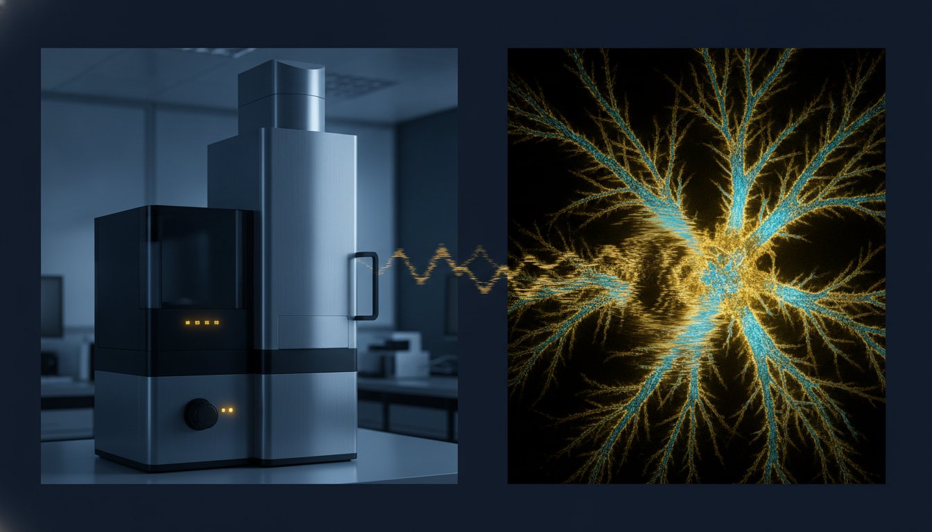
What if the most sophisticated imaging system in your laboratory is currently operating at only sixty percent of its analytical potential due to a misaligned aperture or a degraded tungsten filament? For many industry professionals, the sudden appearance of charging artifacts or persistent astigmatism signifies more than a technical inconvenience; it represents a critical bottleneck that delays essential research and inflates operational costs through emergency service calls. We recognize that maintaining the synergy between vacuum integrity and beam alignment is paramount for achieving the superior standards required in modern materials science.
This professional guide empowers you to master the systematic identification and resolution of hardware failures, offering a definitive approach to troubleshooting common sem operational issues to minimize laboratory downtime. You’ll learn to establish a rigorous protocol for differentiating between simple user error and complex hardware malfunctions that necessitate a professional service contract. By implementing these expert-driven diagnostic narratives, you can restore your Veritas or Cube II systems to peak operational performance while ensuring long-term reliability in your high-resolution imaging workflows.
Key Takeaways
- Establish a methodical approach to vacuum diagnostics and leak detection to ensure the integrity of your electron source and prevent prolonged pump-down cycles.
- Develop a structured diagnostic framework for troubleshooting common sem operational issues, allowing for a clear distinction between rectifiable user errors and critical hardware failures.
- Master the correction of image artifacts, such as astigmatism and sample charging, by optimizing stigmator controls and implementing rigorous sample preparation protocols.
- Identify the specific visual indicators of electronic noise versus mechanical vibration to maintain the crisp imaging standards required for advanced materials analysis.
- Evaluate the financial and operational benefits of professional preventative maintenance as a strategic tool for minimizing unscheduled downtime and preserving the lifecycle of specialized SEM hardware.
Establishing the SEM Operational Baseline
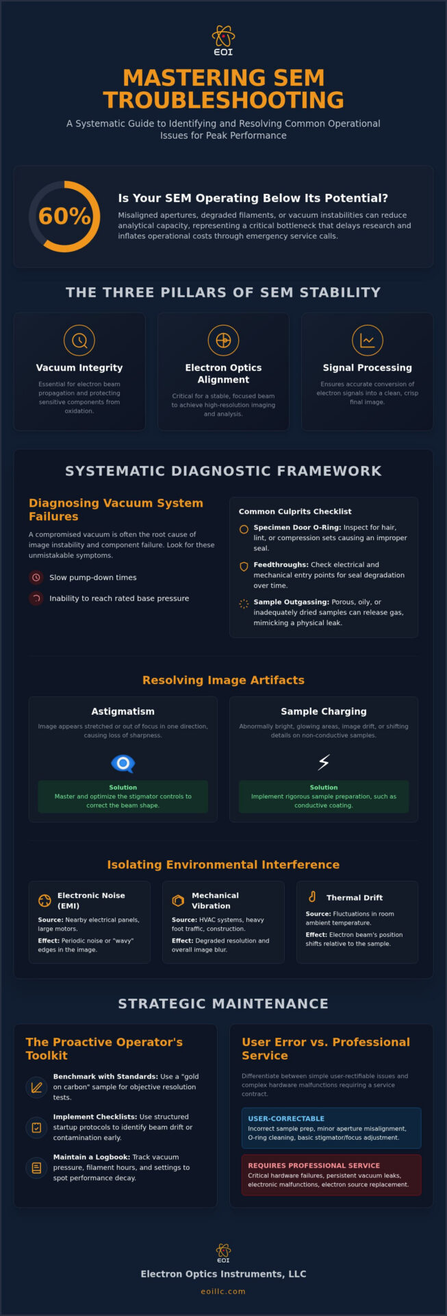
The scanning electron microscope stands as a cornerstone of technical innovation, providing the high-resolution imaging necessary for advanced materials science and industrial quality control. Establishing a rigorous operational baseline is the first prerequisite for any facility aiming for superior standards. It’s essential that industry professionals distinguish these hardware-focused diagnostic protocols from unrelated “Smart Exam Monitor” software issues that often appear in digital documentation. Methodical troubleshooting common sem operational issues requires a deep understanding of the three pillars of stability: vacuum integrity, electron optics alignment, and electronic signal processing. If any of these pillars falter, the entire analytical workflow is compromised.
A reliable benchmarking strategy involves the frequent use of a “gold standard” reference sample, such as gold on carbon or a specialized calibration grid. This practice allows operators to perform objective resolution tests and contrast checks against established factory specifications. By documenting these results, a laboratory can identify subtle performance decay long before it impacts critical production schedules or research outcomes. This meticulousness transforms reactive maintenance into a proactive strategy for long-term reliability.
The Role of the Operator in Performance Monitoring
Technical authority in the lab starts with the operator’s commitment to daily performance monitoring. Implementing structured startup checklists helps identify beam drift or aperture contamination before they cause imaging failure. It’s vital to maintain a comprehensive logbook that tracks vacuum base pressure, filament hours, and aperture settings. This historical data is invaluable when differentiating between routine wear and sudden hardware malfunctions. For those utilizing high-performance systems like the Cube II Benchtop SEM, adhering to a strict monitoring regimen ensures the instrument operates within its designed tolerances, preserving the integrity of every scan.
Environmental Factors Affecting SEM Stability
The vacuum environment is the lifeblood of electron microscopy. Without it, the electron beam cannot propagate, and sensitive components like the tungsten filament or field-emission source will oxidize instantly. Maintaining vacuum integrity is central to troubleshooting common sem operational issues. Systems utilizing field-emission sources require ultra-high vacuum (UHV) levels, often in the range of 10^-7 to 10^-10 Torr, whereas thermionic sources can operate at slightly higher pressures. If the system fails to reach these thresholds, the stability of the entire imaging chain is compromised.
Symptoms of a failing vacuum system are usually unmistakable. Operators often report slow pump-down times or an inability to reach the system’s rated base pressure. When these indicators arise, a systematic approach to leak detection is mandatory. Start with the most frequent point of failure: the specimen chamber door. Inspect the O-ring for hair, lint, or compression sets. If the door is secure, move to the electrical and mechanical feedthroughs. These entry points are susceptible to seal degradation over time, especially in high-throughput laboratories.
Sample outgassing is a frequently overlooked factor in vacuum performance. Porous materials, oils, or inadequately dried biological samples release gas molecules into the chamber, which artificially inflates the pressure. This phenomenon mimics a physical leak but is entirely dependent on the sample’s chemistry. Ensuring that samples are properly desiccated and handled with gloves is a fundamental step in preserving the chamber’s vacuum health.
Mechanical Pump and Turbo Pump Maintenance
The reliability of your primary and secondary pumps determines the system’s uptime. For rotary vane pumps, monitoring oil levels and clarity is vital. Darkened oil suggests thermal degradation or contamination that could lead to backstreaming, where oil vapors migrate back into the clean vacuum chamber. Turbomolecular pumps require even more vigilance. Listen for unusual acoustic signatures or vibrations, which often signal bearing wear. Implementing a routine SEM preventative maintenance program ensures these high-speed components are serviced before a catastrophic failure occurs.
O-Ring Care and Chamber Hygiene
Precision cleaning of Viton O-rings is a technical art. Use only lint-free wipes and avoid solvents that can cause the elastomer to swell or crack. When applying vacuum grease, the “less is more” rule is absolute. Excessive grease traps particulates and contributes to hydrocarbon contamination. For facilities demanding the highest image clarity, utilizing a plasma cleaner to remove residual hydrocarbons from the specimen chamber is a superior standard of care. This meticulousness prevents carbon deposition on the sample surface during high-magnification scans, protecting the optics from long-term degradation.
Resolving Image Artifacts and Beam Instability
Achieving sub-nanometer resolution requires more than just a functional vacuum; it demands absolute control over the electron beam’s geometry and stability. When troubleshooting common sem operational issues, image artifacts often serve as the first diagnostic indicator of an misaligned column or environmental interference. Astigmatism, characterized by a directional blurring that shifts 90 degrees when passing through the focal point, is perhaps the most frequent hurdle. By meticulously adjusting the X and Y stigmator controls, an operator can reshape the elliptical beam into a perfect circle, restoring the crisp edges necessary for high-fidelity imaging. This process is not merely a setting but a continuous calibration that responds to changes in accelerating voltage and working distance.
Distinguishing between mechanical vibration and electronic noise is vital for effective recovery. Mechanical vibration typically manifests as periodic “saw-tooth” edges or jagged lines on vertical features, often originating from nearby heavy machinery or improper stage damping. In contrast, electronic noise appears as a stochastic “snowy” texture across the entire frame. This noise is frequently a result of high detector gain settings or a degrading scintillator. If the image appears to “jump” or shift unexpectedly, the cause is often residual magnetism within the column or localized charging on a non-conductive sample. Optimizing the accelerating voltage and spot size for each specific sample type is the most effective way to balance signal-to-noise ratios against the risk of beam-induced damage.
Filament Saturation and Alignment
Tungsten filament longevity is directly tied to the saturation protocol. Operators should increase the heating current in small, methodical increments until the beam brightness peaks and then levels off. Over-saturation leads to rapid filament thinning, which you can identify by sudden fluctuations in emission current or a noticeable drop in maximum brightness. Once saturated, centering the electron beam within the column apertures is critical. Proper alignment ensures uniform illumination and prevents the “vignetting” effect, where the edges of the field of view appear darker than the center, a common symptom of a poorly centered beam path.
Detectors and Signal Processing Issues
Signal processing failures can lead to “ghosting” or significant lag in the digital display, often indicating a mismatch between the scan rate and the detector’s response time. Troubleshooting Secondary Electron (SE) versus Backscattered Electron (BSE) detector noise requires isolated testing of each signal path to identify if the interference is pre-amplifier or post-amplifier. For laboratories performing elemental analysis, ensuring proper integration with Energy Dispersive Spectroscopy (EDS) systems is paramount. A misaligned beam or incorrect working distance will compromise the X-ray take-off angle, leading to inaccurate stoichiometric data and reduced analytical throughput.
Mitigating Sample Charging and Preparation Errors
Surface charge accumulation remains a persistent hurdle in high-resolution imaging, particularly when analyzing non-conductive polymers or biological specimens. This phenomenon occurs when the rate of electron arrival from the primary beam exceeds the rate of secondary and backscattered electron emission, resulting in a localized negative potential. In the context of troubleshooting common sem operational issues, recognizing visual indicators is essential. Symptoms range from localized bright flares and contrast variations to severe image distortion and “jumping” frames, where the sample appears to shift as the accumulated charge discharges to the nearest ground path.
Effective grounding strategies are the primary defense against these artifacts. Utilizing high-purity carbon tape or conductive silver paint provides a low-resistance path to the microscope stage. For complex geometries, such as those analyzed using the Veritas Series SEM, ensuring that the conductive bridge extends from the top of the sample directly to the aluminum stub is critical. This meticulous preparation prevents the beam from interacting with a floating potential, which otherwise compromises the stability of the scanning raster and the accuracy of the resulting micrograph.
Conductive Coating Techniques
Sputter coating is a standard industrial procedure for transforming non-conductive surfaces into stable analytical targets. The choice of metal is dictated by the required resolution and analytical goals. Gold offers superior conductivity but can exhibit a visible grain structure at magnifications exceeding 50,000x. Platinum or chromium provides a much finer grain for ultra-high-resolution work. Carbon coating is the preferred method for samples destined for elemental analysis, as it minimizes interference with X-ray peaks. Calculating the optimal thickness, typically between 5 and 15 nanometers, is vital to avoid masking fine topographical features. If adhesion fails, it’s often due to residual oils or outgassing, requiring a more rigorous cleaning protocol before the coating process begins.
Alternative Strategies for Non-Conductive Samples
When coating isn’t permissible, such as in forensic analysis or when preserving the sample’s native state is required, alternative strategies must be employed. Utilizing Variable Pressure (VP) or low-vacuum modes allows for the introduction of a controlled gas environment within the chamber. These gas molecules become ionized by the primary beam, effectively neutralizing the surface charge on the specimen. Another effective method for troubleshooting common sem operational issues involves operating at low accelerating voltages, typically below 5kV. At these levels, many materials reach a charge-balance point where the total electron yield equals the primary beam current. For labs facing persistent preparation challenges, consulting with a professional SEM service and support partner can provide the technical insight needed to optimize these difficult imaging sessions.
Strategic Maintenance: When to Call a Professional
While previous sections have addressed the technical nuances of troubleshooting common sem operational issues at the operator level, laboratory managers must recognize the threshold between routine adjustment and professional intervention. Attempting unauthorized repairs on sensitive electron optics or high-voltage power supplies carries significant risk, often leading to irreversible component damage or specialized safety hazards. Strategic maintenance is not merely a reactive necessity; it’s a proactive synergy between internal diligence and external expertise that preserves the instrument’s analytical potential.
The financial benefits of SEM preventative maintenance are well-documented, as regular service intervals significantly reduce the frequency and cost of emergency field repairs. Beyond hardware stability, on-site technical training provided by seasoned engineers can drastically decrease the incidence of operator-induced failures. Electron Optics Instruments leverages over 30 years of industry experience to provide universal support across all major SEM brands, ensuring that your facility maintains international standards of excellence regardless of the specific hardware platform in use.
Understanding SEM Service Contracts
Selecting the appropriate service agreement requires a meticulous evaluation of laboratory throughput and budget constraints. Tiered contracts range from basic preventative visits to comprehensive plans that include full parts and labor coverage. For high-volume industrial environments, a “priority response” clause is essential for minimizing the economic impact of unscheduled downtime. Additionally, a rigorous service history is a critical factor in preserving the long-term resale value of refurbished SEM units, providing a clear audit trail of technical integrity and meticulous care.
Relocation and Re-calibration Requirements
Relocating an electron microscope involves more than physical transport; it requires a professional decommissioning process to secure sensitive internal assemblies against mechanical shock. Post-relocation, the system must undergo a comprehensive re-calibration to verify that it still meets original factory specifications for resolution, magnification accuracy, and beam stability. This methodical approach ensures that the transition between facilities does not compromise your analytical capabilities or data reproducibility. Contact Electron Optics Instruments for expert SEM repair and service.
Advancing Analytical Excellence Through Rigorous Instrument Maintenance
Mastering the intricacies of troubleshooting common sem operational issues is a fundamental prerequisite for any laboratory dedicated to maintaining superior imaging standards and operational continuity. By establishing a robust performance baseline and implementing systematic protocols for vacuum diagnostics and artifact correction, operators can significantly mitigate the impact of unscheduled downtime. This technical meticulousness ensures that advanced systems, such as the Veritas series, consistently operate at their peak analytical potential while preserving the integrity of critical research data.
Electron Optics Instruments reinforces your laboratory’s capabilities through more than 30 years of industry experience and our status as the sole US distributor for EmCraft scanning electron microscopes. We offer comprehensive national service coverage for all major SEM brands, providing the reliability and expertise necessary to navigate complex hardware failures that exceed routine operator adjustments. We invite you to Request a Professional SEM Service Quote or Technical Consultation to secure the longevity and precision of your high-resolution systems. Maintaining the synergy between technical innovation and practical reliability is well within your reach.
Frequently Asked Questions
How do I know if my SEM filament is about to break?
Indicators of imminent filament failure include erratic emission current readings, a significant drop in maximum brightness, or the requirement of higher heating currents to reach saturation. Operators should meticulously monitor the filament’s age in hours as part of their daily log. A sudden “open circuit” error during the startup sequence is the final confirmation of failure. Predicting this point is a vital part of troubleshooting common sem operational issues to prevent downtime during critical imaging sessions.
What is the most common cause of poor SEM image resolution?
Incorrect stigmator adjustment and aperture contamination are the primary culprits for resolution decay in most laboratory settings. If the electron beam isn’t perfectly circular, the resulting astigmatism blurs fine topographical details and creates directional stretching. Additionally, a dirty final aperture can cause beam tailing or irregular charging artifacts. Operators must ensure the optical column is aligned and the working distance is optimized for the specific accelerating voltage to achieve theoretical resolution limits.
Can I fix a vacuum leak in my SEM myself?
Basic leaks at the specimen chamber door or accessible O-rings can often be resolved by the operator through careful cleaning and re-seating of the seals. However, leaks originating from internal feedthroughs, bellows, or high-vacuum valves require professional intervention. Attempting to disassemble vacuum manifolds without specialized diagnostic tools risks introducing further contamination or mechanical damage. If routine maintenance of external seals doesn’t restore base pressure, it’s time to consult a technical authority for advanced leak detection.
Why does my SEM image appear to be “drifting” or moving during capture?
Image drift is frequently caused by thermal instability in the laboratory environment or localized charging on the sample surface. If the microscope’s internal components haven’t reached thermal equilibrium, the column may expand or contract slightly, shifting the beam position relative to the stage. Alternatively, a non-conductive sample that isn’t properly grounded will accumulate charge, creating an electrostatic field that deflects the scanning raster. Ensuring stable ambient temperatures and rigorous grounding protocols is essential for stable imaging.
What are the signs that my SEM column needs professional cleaning?
Persistent image distortion, irregular beam shifts when changing magnification, and uncorrectable astigmatism suggest that the internal column liners or apertures are contaminated with hydrocarbons. As the electron beam interacts with these deposits, they become charged, leading to unpredictable beam deflection and reduced contrast. When standard user-level aperture cleaning fails to restore image clarity, a professional deep-cleaning of the electron optics is necessary to maintain the superior standards required for high-resolution industrial analysis.
How often should preventative maintenance be performed on a benchtop SEM?
Most high-utilization laboratories should schedule preventative maintenance visits every six to twelve months to ensure long-term reliability and accuracy. Even compact systems like the Cube II require regular checks of the vacuum pump oil, cooling systems, and electrical calibrations to prevent drift. This proactive approach is a cornerstone of troubleshooting common sem operational issues, as it identifies wear in mechanical components before they lead to catastrophic hardware failure or expensive, unscheduled emergency repairs.
Why is my sample “charging” even after I have sputter-coated it?
Charging after coating usually indicates an incomplete conductive path or a coating layer that is too thin for the chosen accelerating voltage. If the metal film doesn’t make direct contact with the conductive adhesive or the aluminum stub, the surface remains electrically isolated. Furthermore, high-energy beams can penetrate thin coatings, interacting with the non-conductive bulk material beneath. Re-applying silver paint to the sample edges to ensure a continuous ground path often resolves these persistent failures.
What should I do if the SEM vacuum pump is making a loud grinding noise?
You should immediately vent the system and power down the pump to prevent permanent mechanical seizure or internal damage. Grinding noises typically indicate bearing failure in a turbomolecular pump or a broken vane in a rotary pump. Continued operation under these conditions can lead to catastrophic debris migration into the clean microscope chamber, necessitating a full column overhaul. Contacting a professional service provider for an immediate inspection is the only safe path to system recovery.